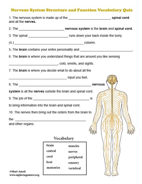
Anatomy And Physiology Nervous System Practice Test
Muz Play
Apr 06, 2025 · 7 min read

Table of Contents
Anatomy and Physiology Nervous System Practice Test: A Comprehensive Review
This comprehensive practice test covers key concepts in the anatomy and physiology of the nervous system. It's designed to help you assess your understanding and identify areas needing further review. Remember, understanding the nervous system requires grasping its intricate structure and complex functions. This test will delve into various aspects, from basic neuron structure to higher-level cognitive processes. Good luck!
Section 1: Neuron Structure and Function
Instructions: Choose the best answer for each multiple-choice question.
1. Which of the following is NOT a part of a neuron?
a) Dendrite b) Axon c) Synapse d) Myelin Sheath e) Nucleus
Answer: C (Synapses are the junctions between neurons, not parts of a single neuron.)
2. What is the primary function of the myelin sheath?
a) To produce neurotransmitters b) To increase the speed of nerve impulse transmission c) To protect the neuron from damage d) To generate action potentials e) Both b and c
Answer: E (Myelin sheaths accelerate nerve impulse transmission and offer a degree of protection.)
3. What type of neuron transmits signals from the central nervous system to muscles or glands?
a) Sensory neuron b) Motor neuron c) Interneuron d) Glial cell e) Neuroglia
Answer: B (Motor neurons carry signals from the CNS to effectors.)
4. The resting membrane potential of a neuron is primarily maintained by:
a) Sodium-potassium pumps b) Diffusion of potassium ions c) Diffusion of sodium ions d) Active transport of chloride ions e) Both a and b
Answer: E (Sodium-potassium pumps actively transport ions, and potassium diffusion contributes significantly to the resting potential.)
5. What event triggers the release of neurotransmitters from the presynaptic neuron?
a) Depolarization of the dendrites b) Hyperpolarization of the axon terminal c) Arrival of an action potential at the axon terminal d) Repolarization of the soma e) Release of calcium ions from the postsynaptic neuron
Answer: C (The action potential reaching the terminal triggers neurotransmitter release.)
Section 2: Central and Peripheral Nervous Systems
Instructions: Answer the following questions in complete sentences.
1. Describe the main functions of the central nervous system (CNS).
The central nervous system, comprising the brain and spinal cord, is responsible for integrating sensory information, processing information, coordinating bodily functions, and generating motor commands. It's the control center for the body's activities. The brain handles higher-level functions like thought, memory, and emotion, while the spinal cord acts as the primary pathway for signals between the brain and the rest of the body.
2. Differentiate between the somatic and autonomic nervous systems. Provide examples of functions controlled by each.
The somatic nervous system (SNS) controls voluntary movements of skeletal muscles. For example, consciously deciding to lift your arm or walk involves the SNS. The autonomic nervous system (ANS), in contrast, regulates involuntary functions such as heart rate, digestion, and breathing. The ANS further divides into the sympathetic (fight-or-flight) and parasympathetic (rest-and-digest) branches, each influencing these involuntary processes in opposing ways.
3. Explain the role of glial cells in the nervous system.
Glial cells, also known as neuroglia, are non-neuronal cells that provide support and protection for neurons. They perform various crucial functions including: providing structural support, supplying nutrients to neurons, insulating axons with myelin, removing waste products, and forming the blood-brain barrier. Different types of glial cells (astrocytes, oligodendrocytes, Schwann cells, microglia, etc.) have specific roles in maintaining the health and function of the nervous system.
4. Describe the protective layers surrounding the brain and spinal cord (meninges).
The brain and spinal cord are encased in three protective layers of connective tissue called meninges: the dura mater (outermost, tough layer), the arachnoid mater (middle, web-like layer containing cerebrospinal fluid), and the pia mater (innermost, delicate layer adhering to the brain and spinal cord). These layers provide cushioning and protection against mechanical injury.
5. What is cerebrospinal fluid (CSF) and what are its functions?
Cerebrospinal fluid (CSF) is a clear, colorless fluid that circulates within the ventricles of the brain, the subarachnoid space (between the arachnoid and pia mater), and the central canal of the spinal cord. Its main functions include cushioning the brain and spinal cord, providing buoyancy to reduce their weight, transporting nutrients and removing waste products, and maintaining a stable chemical environment for the nervous tissue.
Section 3: Brain Regions and Functions
Instructions: Match the brain region in Column A with its primary function in Column B.
Column A Column B
a) Cerebrum 1) Coordinates movement and balance b) Cerebellum 2) Regulates vital functions like breathing and heart rate c) Brainstem 3) Higher-level cognitive functions, sensory processing, and voluntary movement d) Hypothalamus 4) Relays sensory information to the cerebrum e) Thalamus 5) Controls body temperature, hunger, and thirst
Answers: a-3, b-1, c-2, d-5, e-4
Section 4: Sensory Systems and Perception
Instructions: Answer the following questions briefly.
1. What is sensory transduction?
Sensory transduction is the process by which a sensory receptor converts a physical stimulus (light, sound, pressure, etc.) into an electrical signal (receptor potential) that can be transmitted by neurons.
2. Describe the pathway of light through the eye, starting with the cornea and ending with the visual cortex.
Light passes through the cornea, pupil (controlled by the iris), lens (which focuses the light), and then onto the retina. Photoreceptor cells (rods and cones) in the retina convert light into electrical signals, which are then processed by bipolar cells, ganglion cells, and eventually transmitted via the optic nerve to the visual cortex in the occipital lobe for interpretation.
3. Explain the difference between rods and cones in the retina.
Rods are responsible for vision in low-light conditions (night vision), providing low-acuity, black-and-white vision. Cones, on the other hand, mediate high-acuity color vision in bright light conditions.
4. What is the role of the cochlea in hearing?
The cochlea is a spiral-shaped structure in the inner ear that contains hair cells, the sensory receptors for hearing. Sound vibrations transmitted through the middle ear cause fluid movement within the cochlea, bending the hair cells and triggering the generation of nerve impulses that are transmitted to the auditory cortex for sound perception.
5. How do mechanoreceptors contribute to our sense of touch?
Mechanoreceptors are sensory receptors that respond to mechanical pressure or distortion. In the skin, various types of mechanoreceptors detect different types of touch sensations (light touch, pressure, vibration, etc.). These receptors transduce mechanical stimuli into electrical signals that are transmitted to the somatosensory cortex for processing and perception of touch.
Section 5: Higher-Level Brain Functions
Instructions: Answer the following essay question.
1. Discuss the roles of different brain regions in memory formation and retrieval.
Memory formation and retrieval involve intricate interactions between various brain regions. The hippocampus plays a crucial role in the consolidation of new memories, particularly declarative memories (facts and events). It acts as a temporary storage site, transferring memories to other brain regions for long-term storage. The amygdala is involved in processing emotional aspects of memories, particularly those associated with fear or strong emotions. The cerebellum plays a significant role in procedural memory (motor skills and habits). The cerebral cortex, specifically the frontal and temporal lobes, stores long-term memories and contributes to memory retrieval. The prefrontal cortex is important for working memory, allowing us to hold information in mind for short periods and manipulate it. Different types of memory (episodic, semantic, procedural) may involve different neural circuits and brain regions. Damage to specific brain regions can result in selective memory impairments, highlighting the specialized roles of these areas in memory processing.
Section 6: Neurological Disorders
Instructions: Identify the neurological disorder that best matches each description.
1. Progressive neurodegenerative disorder characterized by tremors, rigidity, and bradykinesia.
a) Epilepsy b) Parkinson's disease c) Alzheimer's disease d) Multiple sclerosis
Answer: B
2. Autoimmune disorder that attacks the myelin sheath of neurons.
a) Epilepsy b) Parkinson's disease c) Alzheimer's disease d) Multiple sclerosis
Answer: D
3. Characterized by recurrent seizures.
a) Epilepsy b) Parkinson's disease c) Alzheimer's disease d) Multiple sclerosis
Answer: A
4. Progressive neurodegenerative disorder leading to memory loss, cognitive decline, and behavioral changes.
a) Epilepsy b) Parkinson's disease c) Alzheimer's disease d) Multiple sclerosis
Answer: C
This practice test provides a substantial review of key concepts in the anatomy and physiology of the nervous system. Remember to consult your textbook and class notes for more detailed information. Consistent study and review are key to mastering this complex subject. Good luck with your studies!
Latest Posts
Latest Posts
-
The Image Formed In A Plane Mirror Is
Apr 07, 2025
-
Plants Are Photosynthetic Autotrophs What Does This Mean
Apr 07, 2025
-
Equilibrium And Le Chateliers Principle Lab Answer Key
Apr 07, 2025
-
Bile Is Stored And Concentrated In The
Apr 07, 2025
-
Moment Of Inertia Rod About Center
Apr 07, 2025
Related Post
Thank you for visiting our website which covers about Anatomy And Physiology Nervous System Practice Test . We hope the information provided has been useful to you. Feel free to contact us if you have any questions or need further assistance. See you next time and don't miss to bookmark.
