Collection Of Nerve Cell Bodies In The Pns
Muz Play
Apr 05, 2025 · 7 min read

Table of Contents
Collections of Nerve Cell Bodies in the PNS: A Comprehensive Overview
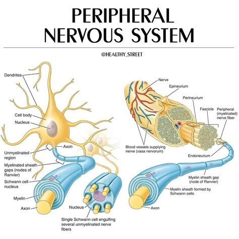
The peripheral nervous system (PNS) is a complex network responsible for connecting the central nervous system (CNS) to the limbs and organs. While the CNS houses the brain and spinal cord, the PNS comprises a vast array of nerves and ganglia. A key component of the PNS architecture are the collections of nerve cell bodies, known as ganglia. Understanding these ganglia is crucial for comprehending the intricate functioning of the PNS and diagnosing various neurological disorders. This article delves deep into the different types of ganglia found in the PNS, their structures, functions, and clinical significance.
Types of Ganglia in the Peripheral Nervous System
Ganglia in the PNS are broadly classified based on their function and the type of neurons they contain:
1. Sensory Ganglia (Posterior Root Ganglia, Dorsal Root Ganglia, Cranial Nerve Ganglia)
These ganglia house the cell bodies of sensory neurons, also known as afferent neurons. These neurons transmit sensory information from the periphery (skin, muscles, organs) to the CNS.
-
Location: Sensory ganglia are located along the dorsal roots of spinal nerves and in association with certain cranial nerves. The spinal sensory ganglia are also known as dorsal root ganglia (DRG) or posterior root ganglia (PRG). Cranial nerve ganglia are named according to the specific cranial nerve they're associated with (e.g., trigeminal ganglion, geniculate ganglion).
-
Structure: Sensory ganglia are typically oval-shaped structures containing pseudounipolar neurons. These neurons have a single process that bifurcates into two branches: one extending peripherally to receive sensory input, and the other extending centrally to synapse with neurons in the CNS. The ganglia are encapsulated by a connective tissue sheath and are richly supplied with blood vessels.
-
Function: The primary function of sensory ganglia is to relay sensory information, including touch, temperature, pain, and proprioception (body position), to the CNS for processing and interpretation. This information is essential for the body's response to its internal and external environment.
-
Clinical Significance: Damage or dysfunction in sensory ganglia can lead to various sensory disturbances, such as numbness, paresthesia (tingling or prickling sensation), pain, and loss of proprioception. These conditions can be caused by infections (e.g., shingles), autoimmune disorders, tumors, and trauma. Conditions like Ramsay Hunt syndrome, characterized by facial paralysis and ear pain, are directly linked to inflammation of the geniculate ganglion.
2. Autonomic Ganglia
These ganglia are crucial components of the autonomic nervous system (ANS), which regulates involuntary functions like heart rate, digestion, and respiration. Autonomic ganglia are further subdivided based on their location within the ANS pathways:
-
Sympathetic Ganglia: These ganglia are part of the sympathetic nervous system, which prepares the body for "fight or flight" responses. They are located in two major chains running alongside the vertebral column (paravertebral ganglia) and in prevertebral plexuses (e.g., celiac plexus, mesenteric plexus).
-
Structure: Sympathetic ganglia are typically interconnected and form complex networks. They contain multi-polar neurons that receive input from preganglionic sympathetic neurons originating in the spinal cord.
-
Function: Sympathetic ganglia mediate the release of neurotransmitters (primarily norepinephrine) that influence target organs, increasing heart rate, blood pressure, and respiration, while diverting blood flow away from non-essential organs.
-
Clinical Significance: Dysfunction in sympathetic ganglia can manifest in a variety of ways, including orthostatic hypotension (low blood pressure upon standing), impaired thermoregulation, and gastrointestinal problems. Damage can result from trauma, surgery, or autoimmune diseases like autoimmune autonomic ganglionopathy.
-
-
Parasympathetic Ganglia: These ganglia belong to the parasympathetic nervous system, which promotes "rest and digest" functions. They are located near or within the target organs they innervate.
-
Structure: Parasympathetic ganglia are smaller and less numerous than sympathetic ganglia. They contain multipolar neurons that receive input from preganglionic parasympathetic neurons originating in the brainstem and sacral spinal cord.
-
Function: Parasympathetic ganglia release acetylcholine, which slows heart rate, stimulates digestion, and promotes other restorative functions.
-
Clinical Significance: Damage to parasympathetic ganglia can cause difficulties with bowel and bladder control, altered heart rate response, and dry mouth (xerostomia). Conditions like Shy-Drager syndrome involve widespread autonomic nervous system dysfunction impacting parasympathetic ganglia.
-
Neurotransmitters and Receptors in Ganglia
The functionality of ganglia is heavily dependent on the neurotransmitters released by preganglionic neurons and the receptors present on postganglionic neurons.
-
Acetylcholine: This is the primary neurotransmitter released by preganglionic neurons in both the sympathetic and parasympathetic nervous systems. It binds to nicotinic acetylcholine receptors (nAChRs) on postganglionic neurons.
-
Norepinephrine: This is the primary neurotransmitter released by postganglionic sympathetic neurons. It binds to adrenergic receptors (α and β receptors) on target organs.
-
Acetylcholine: In the parasympathetic nervous system, postganglionic neurons also release acetylcholine, which binds to muscarinic acetylcholine receptors (mAChRs) on target organs.
Understanding the specific neurotransmitters and receptors involved is critical for the development of pharmaceuticals targeting specific aspects of the autonomic nervous system. For instance, drugs that block certain adrenergic receptors are used to treat hypertension, while others targeting mAChRs are used in the treatment of asthma.
Microscopic Structure of Ganglia
At a microscopic level, ganglia are highly organized structures:
-
Neuron Cell Bodies: The most prominent feature is the presence of numerous neuron cell bodies, each with its nucleus and surrounding cytoplasm. In sensory ganglia, these are pseudounipolar neurons, while in autonomic ganglia they are multipolar neurons.
-
Satellite Cells (Glial Cells): These glial cells surround and support the neuron cell bodies. They provide trophic support, insulation, and protection. In sensory ganglia, satellite cells are crucial for maintaining the microenvironment around the neuron cell bodies.
-
Connective Tissue: Ganglia are enclosed within a connective tissue capsule that provides structural support and protection. This capsule extends into the ganglion, forming septa that divide the ganglion into lobules. Blood vessels run within the connective tissue, supplying oxygen and nutrients to the neurons and satellite cells.
-
Axons and Dendrites: Axons and dendrites extend from the neuron cell bodies, forming the afferent and efferent pathways of the ganglia. Myelin sheaths may be present on axons depending on the type of neuron and its location.
Clinical Relevance and Diseases Affecting Ganglia
Various diseases and conditions can affect the ganglia of the PNS, leading to a range of symptoms:
-
Shingles (Herpes Zoster): Caused by the varicella-zoster virus, this infection affects sensory ganglia, typically resulting in painful skin rashes along the dermatomes (areas of skin innervated by a specific sensory nerve).
-
Guillain-Barré Syndrome (GBS): This autoimmune disorder attacks the myelin sheaths of peripheral nerves, potentially including axons within ganglia. Symptoms include progressive muscle weakness and paralysis.
-
Amyloidosis: Abnormal protein deposits (amyloid) can accumulate in ganglia, impairing neuronal function and leading to various symptoms depending on the affected ganglia.
-
Neuroblastoma: This malignant tumor originates from neuroblasts, immature nerve cells, frequently found in the adrenal glands or sympathetic ganglia.
-
Ganglioneuroma: A benign tumor arising from mature nerve cells in ganglia.
-
Multiple System Atrophy (MSA): A rare, progressive neurological disorder that affects the autonomic nervous system, often leading to significant dysfunction of ganglia.
Early diagnosis and appropriate management are crucial in minimizing the impact of these diseases on patients' lives.
Further Research and Future Directions
Ongoing research continues to unravel the complexities of PNS ganglia, focusing on:
-
Development and Regeneration: Studies aim to understand how ganglia develop and how to promote regeneration after injury or disease.
-
Role in Pain Perception: Research investigates the mechanisms involved in pain signaling within sensory ganglia and the potential for developing novel pain management strategies.
-
Autonomic Dysfunction: Investigations focus on identifying the underlying mechanisms of autonomic dysfunction and developing effective treatments for conditions affecting autonomic ganglia.
-
Genetic Basis of Ganglia-Related Diseases: Genetic studies explore the role of inherited factors in the development and susceptibility to diseases affecting ganglia.
The study of ganglia is a dynamic field with immense potential for advancing our understanding of the PNS and developing innovative therapeutic interventions for a range of neurological disorders. Continued research will be vital in addressing the clinical challenges posed by ganglia-related diseases.
Latest Posts
Latest Posts
-
Chemistry Matter And Change Textbook Pdf
Apr 06, 2025
-
What Is Two Types Of Reproduction
Apr 06, 2025
-
7 1 Solve Linear Systems By Graphing
Apr 06, 2025
-
Are Lewis Structures Only For Covalent Bonds
Apr 06, 2025
-
An Acid Which Ionizes Completely In Water Is A
Apr 06, 2025
Related Post
Thank you for visiting our website which covers about Collection Of Nerve Cell Bodies In The Pns . We hope the information provided has been useful to you. Feel free to contact us if you have any questions or need further assistance. See you next time and don't miss to bookmark.
