Dissection Of Cat Veins And Arteries
Muz Play
Apr 07, 2025 · 6 min read
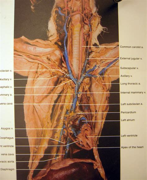
Table of Contents
Dissection of Cat Veins and Arteries: A Comprehensive Guide
Understanding the circulatory system is fundamental to veterinary and human anatomy. Dissection of a feline circulatory system provides a hands-on approach to learning the intricate network of veins and arteries. This detailed guide will walk you through the process, highlighting key anatomical structures and providing essential tips for a successful dissection. Note: This guide is intended for educational purposes and should only be performed under the supervision of a qualified instructor with proper ethical considerations and adherence to all relevant regulations regarding animal usage in education.
Preparing for the Dissection
Before beginning, ensure you have the necessary materials and understand the ethical implications. Obtaining a feline specimen ethically is crucial. Many educational institutions have established protocols for procuring specimens for anatomical study, ensuring compliance with all relevant regulations. Always prioritize ethical sourcing and respect for the animal.
Materials You Will Need:
- Dissecting Tray: A sturdy tray to hold the specimen and prevent spills.
- Dissecting Kit: This should include scalpels (various sizes), forceps (with teeth and without), scissors (sharp and blunt-tipped), probes, and pins.
- Gloves: Essential for hygiene and safety.
- Apron: To protect your clothing.
- Eye Protection: Safety glasses are highly recommended.
- Preserved Feline Specimen: Ideally, a specimen that has been properly preserved to maintain anatomical integrity.
- Dissecting Guide: An anatomical atlas of the feline circulatory system will be invaluable.
- Camera (Optional): To document your findings and progress.
- Waste Disposal: A container for proper disposal of used materials.
External Anatomy: Initial Observations
Begin by carefully examining the external anatomy of the cat. Note the location of the major blood vessels visible on the surface. These will serve as landmarks for your dissection. Look for:
- Jugular Veins: These are prominent veins located on either side of the neck, carrying deoxygenated blood from the head and neck back to the heart.
- Femoral Artery and Vein: These vessels are found in the groin region of the hind legs, easily identifiable by their size and proximity.
- Saphenous Vein: This superficial vein runs along the medial aspect of the hind leg.
- Cephalic Vein: Located on the anterior (front) aspect of the forelimb.
Take detailed notes and sketches of your initial observations. This will be crucial for later identification of internal structures.
Internal Anatomy: A Step-by-Step Dissection
1. Incision: Begin by making a midline incision through the skin along the ventral (belly) surface of the cat, extending from the lower jaw to the pubic region. Use sharp scissors to carefully cut through the skin, avoiding any unnecessary damage to underlying tissues.
2. Muscle Layers: Once the skin is opened, you’ll encounter several layers of muscle. Carefully separate these layers using blunt dissection techniques (avoiding sharp instruments where possible). This minimizes damage to the underlying blood vessels.
3. Locating Major Blood Vessels: As you proceed through the muscle layers, you will begin to identify major arteries and veins.
- Aorta: The main artery leaving the heart, carrying oxygenated blood to the rest of the body. Locate its branching pattern, carefully observing the arteries supplying the different organs. Pay close attention to the brachiocephalic artery, which branches off the aorta and supplies the head and forelimbs; the carotid arteries, which supply the head; and the subclavian arteries, which supply the forelimbs.
- Vena Cava: The large vein returning deoxygenated blood to the heart from the body. Note its location and the veins draining into it, such as the jugular veins, iliac veins, and renal veins.
- Pulmonary Artery and Veins: These vessels are located close to the heart. The pulmonary artery carries deoxygenated blood to the lungs, while the pulmonary veins return oxygenated blood to the heart.
4. Tracing Blood Vessels: Use blunt probes and forceps to gently lift and separate tissues, carefully tracing the paths of the major blood vessels. Note their branching patterns, size, and relationships to surrounding structures.
5. Detailed Examination of Specific Regions:
- Heart: Carefully expose the heart, observing its size, shape, and location within the thoracic cavity. Note the location of the great vessels entering and leaving the heart.
- Liver: Examine the hepatic portal vein and its branches, understanding its role in processing nutrients absorbed from the digestive system.
- Kidneys: Observe the renal artery and vein, crucial for filtering blood and producing urine.
- Spleen: Note the splenic artery and vein.
- Brain: (This requires additional care and specialized tools) Carefully dissect the skull to expose the circle of Willis, a vital network of arteries supplying blood to the brain.
6. Documentation: Throughout the dissection, take detailed notes and photographs to record your findings. Clearly label all structures and their relationships to surrounding tissues.
Understanding the Feline Circulatory System
The feline circulatory system, like that of other mammals, is a closed system. Blood is propelled through the heart, arteries, capillaries, and veins in a continuous circuit.
Arteries: Carry oxygenated blood away from the heart (except the pulmonary artery). They have thick, elastic walls to withstand the high pressure of blood pumped by the heart. They branch into smaller arterioles and eventually capillaries.
Veins: Carry deoxygenated blood back towards the heart (except the pulmonary veins). They have thinner walls than arteries and often contain valves to prevent backflow of blood. Venules merge to form larger veins.
Capillaries: Microscopic vessels connecting arteries and veins. They have extremely thin walls, facilitating the exchange of gases, nutrients, and waste products between blood and tissues.
Key Differences Between Feline and Human Circulatory Systems
While the general structure of the circulatory system is similar across mammals, there are some key differences between cats and humans:
- Size and Branching Patterns: The specific size and branching patterns of arteries and veins may vary slightly between species.
- Anatomical Variations: Minor anatomical variations can occur even within the same species.
- Physiological Differences: The metabolic rate and overall physiology of a cat differ from that of a human, impacting the circulatory system's functioning.
Safety Precautions and Ethical Considerations
- Sterile Technique: Maintain a sterile environment as much as possible to prevent infection.
- Sharp Instrument Handling: Always use sharp instruments with care to avoid injury.
- Waste Disposal: Dispose of all biological materials properly according to established guidelines.
- Ethical Sourcing: Ensure your specimen was obtained ethically and legally.
Conclusion
Dissection of a cat's veins and arteries offers a unique and invaluable learning experience. By meticulously following the steps outlined in this guide, and with proper preparation and ethical considerations, you can gain a deep understanding of the feline circulatory system and its crucial role in maintaining life. Remember to always prioritize safety, respect, and proper disposal techniques. This comprehensive dissection will provide a strong foundation for further studies in comparative anatomy, physiology, and veterinary medicine. The experience of hands-on learning will solidify your understanding of the complex network of vessels that sustain life within the feline body. This detailed approach enhances the educational value far beyond simply reading about the structures; it allows for a direct, tactile understanding of their three-dimensional arrangement and their relationships with other organs and tissues.
Latest Posts
Latest Posts
-
What Makes A Cell A Target Of A Particular Hormone
Apr 10, 2025
-
What Is A Period Number On The Periodic Table
Apr 10, 2025
-
How Does The Inorganic Portion Of Soil Form
Apr 10, 2025
-
What Are Intermediates In A Reaction
Apr 10, 2025
-
Is Sodium Most Likekey To Become A Cations
Apr 10, 2025
Related Post
Thank you for visiting our website which covers about Dissection Of Cat Veins And Arteries . We hope the information provided has been useful to you. Feel free to contact us if you have any questions or need further assistance. See you next time and don't miss to bookmark.