Functional Microscopic Anatomy Of The Kidney
Muz Play
Apr 06, 2025 · 6 min read
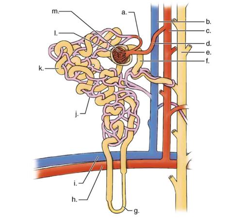
Table of Contents
Functional Microscopic Anatomy of the Kidney
The kidney, a vital organ in the urinary system, plays a crucial role in maintaining homeostasis by regulating fluid balance, electrolyte levels, and blood pressure, while also eliminating metabolic waste products. Understanding its intricate microscopic anatomy is key to comprehending its complex functional capabilities. This article delves into the functional microscopic anatomy of the kidney, exploring the nephron, its components, and the processes involved in urine formation.
The Nephron: The Functional Unit of the Kidney
The basic functional unit of the kidney is the nephron, numbering approximately one million in each kidney. Each nephron consists of two main parts: the renal corpuscle and the renal tubule. The intricate interplay between these structures allows for the precise filtration, reabsorption, and secretion processes that ultimately produce urine.
Renal Corpuscle: Filtration's First Line of Defense
The renal corpuscle, located in the cortex of the kidney, is responsible for the initial filtration of blood. It comprises two key structures:
-
Glomerulus: A network of specialized capillaries, the glomerulus receives blood from an afferent arteriole and is characterized by its high permeability. This high permeability is crucial for efficient filtration. The fenestrated endothelium of the glomerular capillaries allows for the passage of water and small solutes while largely excluding larger proteins and blood cells.
-
Bowman's Capsule: A double-walled epithelial cup surrounding the glomerulus, Bowman's capsule collects the filtrate produced by the glomerulus. The visceral layer of Bowman's capsule, made up of specialized podocytes, plays a crucial role in regulating filtration. Podocytes possess foot-like processes called pedicels that interdigitate, creating filtration slits that further refine the filtration process. The filtration slits are spanned by a specialized membrane, the slit diaphragm, that restricts the passage of even smaller proteins.
The interplay between the fenestrated endothelium, the glomerular basement membrane, and the filtration slits of the podocytes creates a selective filter, allowing for the efficient removal of waste products while retaining essential proteins and blood cells. This process, known as glomerular filtration, is driven by the hydrostatic pressure difference between the glomerular capillaries and Bowman's capsule.
Renal Tubule: Fine-tuning the Filtrate
The filtrate produced by the glomerulus then enters the renal tubule, a long, convoluted tube responsible for modifying the filtrate through reabsorption and secretion. The renal tubule can be divided into several segments, each with distinct characteristics and functions:
-
Proximal Convoluted Tubule (PCT): The PCT is the longest segment of the renal tubule and is characterized by its brush border, a dense collection of microvilli on the luminal surface. This brush border significantly increases the surface area available for reabsorption. The PCT is responsible for the bulk reabsorption of water, glucose, amino acids, ions (sodium, potassium, chloride, bicarbonate), and other essential nutrients. It also plays a role in secretion, removing certain substances like hydrogen ions and drugs from the blood into the tubular fluid.
-
Loop of Henle: This U-shaped structure extends into the medulla of the kidney and plays a vital role in concentrating the urine. The descending limb of the Loop of Henle is highly permeable to water but relatively impermeable to solutes. The ascending limb, in contrast, is impermeable to water but actively transports sodium, potassium, and chloride ions out of the tubular fluid, contributing to the establishment of a concentration gradient in the medulla. This countercurrent mechanism is crucial for water reabsorption in the collecting duct.
-
Distal Convoluted Tubule (DCT): The DCT is shorter than the PCT and has a less prominent brush border. It plays a critical role in regulating potassium and calcium balance, as well as fine-tuning sodium reabsorption. Hormones such as aldosterone and parathyroid hormone influence ion transport in the DCT. The DCT also participates in acid-base balance by secreting hydrogen ions.
-
Connecting Tubule and Collecting Duct: These segments receive filtrate from multiple nephrons and play a crucial role in final urine concentration and composition. The collecting ducts run through the medulla, passing through the varying osmotic gradients established by the Loop of Henle. Hormones like antidiuretic hormone (ADH) influence water permeability in the collecting duct, regulating water reabsorption and urine concentration. The final urine is then transported to the renal pelvis and subsequently to the ureter.
Microscopic Processes: A Detailed Look
The functional microscopic anatomy of the kidney allows for several key physiological processes:
1. Glomerular Filtration: The Initial Step
Glomerular filtration is the first step in urine formation and is driven by the hydrostatic pressure difference across the glomerular capillaries. The filtration rate is influenced by several factors, including glomerular capillary pressure, Bowman's capsule hydrostatic pressure, and the oncotic pressure of the glomerular capillaries. The glomerular filtration rate (GFR) is a crucial indicator of kidney function.
2. Tubular Reabsorption: Reclaiming Essentials
Tubular reabsorption involves the transport of essential substances from the filtrate back into the blood. This process is highly selective and occurs in different segments of the renal tubule, primarily the PCT. Different mechanisms, including passive diffusion, facilitated diffusion, active transport, and secondary active transport, are employed to reabsorb various solutes and water.
3. Tubular Secretion: Active Removal of Waste
Tubular secretion is the process by which substances are transported from the blood into the filtrate. This process is important for eliminating waste products, regulating blood pH, and controlling the levels of certain ions. Secretion plays a vital role in the elimination of drugs and toxins.
4. Urine Concentration: Fine-tuning the Output
Urine concentration is a crucial function of the kidney, achieved through the countercurrent mechanism in the Loop of Henle and the action of ADH in the collecting duct. ADH increases the permeability of the collecting duct to water, allowing for greater water reabsorption and the production of concentrated urine.
Clinical Significance: Microscopic Insights into Disease
Understanding the functional microscopic anatomy of the kidney is crucial in diagnosing and managing various renal diseases. Microscopic changes in the nephrons can provide valuable insights into the pathophysiology of kidney disorders. For example:
-
Glomerulonephritis: Inflammation of the glomeruli can lead to impaired filtration and proteinuria (protein in the urine). Microscopic examination of the glomeruli reveals characteristic changes associated with the specific type of glomerulonephritis.
-
Tubular disorders: Damage to the renal tubules can impair reabsorption and secretion, leading to electrolyte imbalances and other complications. Microscopic examination of the tubules can reveal signs of injury or dysfunction.
-
Polycystic kidney disease: This genetic disorder involves the development of numerous cysts in the kidneys, eventually leading to renal failure. Microscopic examination reveals the presence of these cysts within the renal parenchyma.
Conclusion
The functional microscopic anatomy of the kidney is remarkably complex, reflecting its crucial role in maintaining homeostasis. The intricate interplay between the nephron's components, the precise mechanisms of filtration, reabsorption, and secretion, and the influence of hormones all contribute to the kidney's ability to maintain fluid and electrolyte balance, eliminate waste products, and regulate blood pressure. A thorough understanding of this intricate structure and its functions is essential not only for appreciating the marvel of physiological processes but also for diagnosing and managing a wide range of renal diseases. Further research into the microscopic details of renal physiology continues to deepen our understanding and improve clinical practice. This knowledge is critical for advancements in nephrology, contributing to better treatments and improved patient outcomes. The complexity and precision of the kidney's microscopic architecture highlight its vital role in maintaining overall health and well-being.
Latest Posts
Latest Posts
-
Microscopic Anatomy Of Skeletal Muscle Worksheet Answers
Apr 09, 2025
-
Regents Biology Genetics Practice 3 Blood Type Genetics Answer Key
Apr 09, 2025
-
Electric Field Of An Infinite Plane
Apr 09, 2025
-
What Is The Special Property Of Water
Apr 09, 2025
-
What Is A Titrant In Chemistry
Apr 09, 2025
Related Post
Thank you for visiting our website which covers about Functional Microscopic Anatomy Of The Kidney . We hope the information provided has been useful to you. Feel free to contact us if you have any questions or need further assistance. See you next time and don't miss to bookmark.