How To Prepare A Wet Mount Microscope Slide
Muz Play
Apr 03, 2025 · 6 min read

Table of Contents
How to Prepare a Wet Mount Microscope Slide: A Comprehensive Guide
Preparing a wet mount microscope slide is a fundamental skill in biology and microscopy. This technique allows you to observe living organisms and specimens in their natural, aqueous environment, revealing details that might be lost through other preparation methods like staining or sectioning. While seemingly simple, mastering the technique requires attention to detail to avoid artifacts and ensure clear, high-quality observations. This comprehensive guide will walk you through the process step-by-step, addressing common challenges and offering tips for optimal results.
Understanding the Wet Mount Technique
A wet mount involves suspending a specimen in a drop of liquid (typically water, but other solutions may be used) on a microscope slide and then covering it with a coverslip. This creates a temporary, sealed environment for observation. The liquid medium helps to keep the specimen moist and prevents it from drying out under the microscope's light source. The coverslip flattens the specimen, providing a clearer image, and protects the objective lens from potential damage.
Advantages of Wet Mounts:
- Observation of Living Organisms: Wet mounts are ideal for observing living microorganisms like bacteria, protozoa, algae, and small invertebrates.
- Simplicity and Speed: They are relatively quick and easy to prepare, requiring minimal equipment.
- Reversibility: The preparation is temporary, allowing for easy specimen retrieval or re-preparation if needed.
- Cost-Effectiveness: Wet mounts require minimal materials, making them a budget-friendly option.
Disadvantages of Wet Mounts:
- Short-Term Stability: The specimen can dry out quickly, limiting observation time.
- Limited Control: It can be challenging to control the position and orientation of the specimen.
- Potential for Artifacts: Air bubbles can interfere with viewing, and the pressure from the coverslip can distort delicate specimens.
- Magnification Limitations: The thickness of the water droplet can limit the use of higher magnification objectives.
Materials Required for Wet Mount Preparation
Before beginning, ensure you have all the necessary materials gathered. This organized approach will prevent interruptions and ensure a smooth preparation process. These materials are readily available in most biology laboratories or can be easily sourced online.
- Microscope Slides: Clean, standard microscope slides are essential. Avoid using slides with scratches or imperfections, as these can interfere with observation.
- Coverslips: Thin, square or circular coverslips are used to cover the specimen and liquid droplet. Their thinness ensures that the objective lens can get sufficiently close to the specimen.
- Specimen: The organism or material you wish to observe under the microscope.
- Distilled Water or Appropriate Mounting Medium: Using distilled or deionized water minimizes the risk of introducing contaminants. Other liquids, such as saline solution or a specific stain, may be appropriate depending on the specimen.
- Dropper or Pipette: Accurate dispensing of the mounting medium is crucial to avoid excess liquid that might overflow or cause artifacts.
- Dissecting Needle or Forceps: These are used to carefully handle delicate specimens and position them appropriately on the slide.
- Lens Paper: Use lint-free lens paper to clean microscope slides and coverslips. Fingerprints and dust can obscure the view.
- Microscope: Obviously, you'll need a microscope to view your prepared slide!
Step-by-Step Guide to Preparing a Wet Mount
Follow these steps carefully to ensure a high-quality wet mount slide preparation. Remember, practice makes perfect!
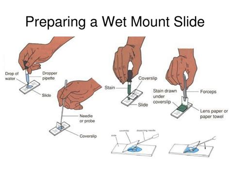
Step 1: Prepare the Slide
Clean the microscope slide thoroughly using lens paper. Any dust particles or fingerprints will be magnified significantly under the microscope and will compromise the quality of your observation.
Step 2: Add a Drop of Mounting Medium
Using a dropper or pipette, place a small drop of distilled water or the appropriate mounting medium in the center of the clean microscope slide. The droplet should be small enough to be fully covered by the coverslip, but large enough to adequately suspend the specimen. Too much liquid will overflow; too little will not properly suspend the specimen.
Step 3: Mount the Specimen
Carefully use a dissecting needle or forceps to place the specimen into the drop of mounting medium. Try to center the specimen for optimal viewing. Gently manipulate the specimen to position it as desired. Avoid pressing too hard, as this could damage delicate specimens.
Step 4: Apply the Coverslip
This step is crucial for avoiding air bubbles and achieving a smooth, even preparation. Hold the coverslip at a 45-degree angle to the slide and carefully lower it onto the droplet. This technique minimizes air bubble entrapment. If air bubbles are present, gently tap the coverslip with the tip of a pencil eraser. This can often help to dislodge the bubbles, though some may be unavoidable.
Step 5: Remove Excess Liquid
If excess liquid has seeped out from under the coverslip, gently blot it away using a piece of absorbent paper or a tissue. Be careful not to disturb the coverslip.
Step 6: Observe Under the Microscope
Place the prepared slide onto the stage of your microscope. Begin with a lower magnification objective (e.g., 4x or 10x) to locate the specimen and then gradually increase magnification to obtain the desired level of detail. Adjust the focus using the coarse and fine focus knobs.
Troubleshooting Common Wet Mount Problems
Even with careful preparation, problems can arise. Here's how to address some common challenges:
- Air Bubbles: As mentioned earlier, lowering the coverslip at an angle minimizes air bubble formation. If bubbles are unavoidable, try gently tapping the coverslip or preparing a new slide.
- Specimen Too Dry: If the specimen is drying out too quickly, add a small ring of petroleum jelly to the edges of the coverslip to create a seal.
- Specimen Too Thick: If the specimen is too thick for proper observation, attempt to dissect or section it to make it thinner.
- Specimen Moving Too Much: For highly mobile organisms, consider using a slightly thicker mounting medium or a slower-moving specimen.
- Coverslip Slipping: Make sure there is sufficient liquid for the coverslip to adhere. If the coverslip is slipping, try adding more mounting medium, though be careful to avoid overflowing.
- Poor Contrast: If the specimen is difficult to see, consider using a contrasting stain that is suitable for living organisms. However, be aware that many stains kill the organisms.
Advanced Wet Mount Techniques
The basic wet mount technique can be adapted for various applications. Here are some advanced considerations:
- Using Stains: While many stains are harmful to live organisms, some vital stains can be used to enhance contrast without killing the specimen.
- Using Specialized Mounting Media: Different mounting media have varying refractive indexes and other properties, making them suitable for different applications. Some media have higher viscosity, making them useful for delicate or moving specimens.
- Creating Hanging Drop Slides: This variation is useful for examining highly mobile microorganisms. A drop of the specimen is suspended from the coverslip, which is then inverted over a well slide, creating a hanging drop.
- Using Chamber Slides: These specialized slides contain wells, providing a contained space for the wet mount, minimizing evaporation and simplifying the process.
Conclusion: Mastering the Wet Mount
Mastering the wet mount technique is a cornerstone of microscopy. While simple in principle, it requires practice and attention to detail to produce high-quality slides. By carefully following these steps and troubleshooting common issues, you can confidently prepare wet mounts for a variety of observations, unlocking a deeper understanding of the microscopic world. Remember, the goal is to produce a clear image of your specimen with minimal artifacts. With persistence and practice, you'll become proficient in preparing effective and informative wet mounts. Happy microscoping!
Latest Posts
Latest Posts
-
Determine The Oxidation State Of Each Species
Apr 04, 2025
-
How To Find The Domain Of A Multivariable Function
Apr 04, 2025
-
A Compound With Two Chirality Centers
Apr 04, 2025
-
Secretion Occurs When Substances Pass From The
Apr 04, 2025
-
How Many Lobes Does The Frogs Liver Have
Apr 04, 2025
Related Post
Thank you for visiting our website which covers about How To Prepare A Wet Mount Microscope Slide . We hope the information provided has been useful to you. Feel free to contact us if you have any questions or need further assistance. See you next time and don't miss to bookmark.
