Label The Arteries Of The Right Lower Limb.
Muz Play
Mar 18, 2025 · 7 min read
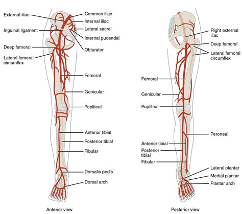
Table of Contents
Label the Arteries of the Right Lower Limb: A Comprehensive Guide
Understanding the arterial supply to the lower limb is crucial for medical professionals, students, and anyone interested in human anatomy. This detailed guide will walk you through the arteries of the right lower limb, providing a comprehensive overview of their branching patterns, locations, and clinical significance. We'll use clear and concise language, supplemented with imagined visuals to aid your understanding. This guide prioritizes accuracy and clarity to ensure you develop a strong understanding of this complex system.
The Major Arteries of the Right Lower Limb: An Overview
The arterial supply to the right lower limb originates from the external iliac artery, which continues as the femoral artery once it passes under the inguinal ligament. This artery, the main vessel of the thigh, gives rise to several crucial branches before continuing its descent. The femoral artery then transitions into the popliteal artery behind the knee, further branching to supply the leg and foot. Let's delve deeper into each section.
1. The Femoral Artery and its Branches: Powering the Thigh
The femoral artery, a continuation of the external iliac artery, is the primary artery supplying the thigh. Imagine it as the main highway, with various smaller roads branching off to deliver blood to different areas. Its key branches include:
-
Superficial Epigastric Artery: A small artery supplying the lower abdominal wall. Think of this as a small side road supplying a small neighborhood.
-
Superficial Circumflex Iliac Artery: This artery runs along the iliac crest, providing blood to the lower abdominal wall and the iliac region. It's like a slightly larger road serving a larger area.
-
Superficial External Pudendal Artery: Supplying blood to the external genitalia. This is a smaller road leading to a more specific location.
-
Deep External Pudendal Artery: Also supplies the external genitalia, offering an alternative route.
-
Deep Femoral Artery (Profunda Femoris): This is the most significant branch of the femoral artery. Consider it a major highway branching off the main one. It further subdivides into:
- Medial Circumflex Femoral Artery: Supplies blood to the head of the femur and surrounding muscles. Think of this as a significant road branching from the major highway to a large area.
- Lateral Circumflex Femoral Artery: Supplies blood to the muscles on the lateral side of the thigh. Similar to the medial circumflex, this is also a critical artery.
- Perforating Arteries: Three to four perforating arteries pierce the adductor magnus muscle, providing blood to the posterior compartment of the thigh. These represent connecting roads between the main artery and the posterior aspect of the thigh.
These branches ensure comprehensive blood supply to all the muscles, bones, and skin of the thigh. Understanding their precise locations is critical for surgical procedures and diagnosis of vascular conditions.
2. The Popliteal Artery: The Knee's Lifeline
As the femoral artery passes behind the knee, it becomes the popliteal artery. This artery, nestled deep within the popliteal fossa (the hollow at the back of the knee), is crucial for supplying blood to the knee joint and the leg. Its branches are vital for the leg's intricate network of muscles and tissues. Key branches of the popliteal artery include:
- Genicular Arteries: These arteries surround the knee joint, providing crucial blood supply for its stability and function. They are like several smaller roads specifically servicing the knee area, ensuring adequate blood flow to the joint. Superior, middle and inferior genicular arteries contribute to this complex network.
The popliteal artery further divides into the anterior and posterior tibial arteries, continuing the blood supply down the leg.
3. The Anterior Tibial Artery: Supplying the Front of the Leg
The anterior tibial artery emerges from the distal end of the popliteal artery, passing between the tibia and fibula. It's the principal artery supplying the anterior compartment of the leg, encompassing the muscles responsible for dorsiflexion of the foot and extension of the toes. Key branches include:
-
Recurrent Tibial Artery: This smaller artery provides additional blood supply to the knee joint.
-
Anterior Tibial Arterial Branches: These smaller arteries supply blood to the anterior leg muscles. Think of them as smaller roads branching off the main highway to supply various buildings (muscles) in that region.
The anterior tibial artery eventually becomes the dorsal pedal artery at the ankle, supplying the dorsum of the foot.
4. The Posterior Tibial Artery: Nourishing the Back of the Leg
The posterior tibial artery, another branch of the popliteal artery, runs down the back of the leg, nestled deep within the posterior compartment. It supplies the posterior leg muscles responsible for plantarflexion of the foot and flexion of the toes. Key branches include:
-
Peroneal Artery (Fibular Artery): This significant branch runs along the fibula, supplying blood to the lateral side of the leg. Imagine this as a major side road supplying a significant portion of the leg’s posterior compartment.
-
Posterior Tibial Arterial Branches: These smaller branches supply the muscles of the posterior leg. They are the smaller roads that feed the muscles, ensuring all parts of the leg receive sufficient blood.
The posterior tibial artery eventually branches into the medial and lateral plantar arteries at the ankle, supplying the sole of the foot.
5. The Medial and Lateral Plantar Arteries: Providing Blood to the Foot's Sole
The medial and lateral plantar arteries, terminal branches of the posterior tibial artery, form the plantar arch, a critical arterial structure in the sole of the foot. This arch supplies blood to the intrinsic muscles of the foot, providing the necessary oxygen and nutrients for movement and function.
6. The Dorsal Pedal Artery: Circulation to the Top of the Foot
The anterior tibial artery transitions into the dorsal pedal artery, which runs along the dorsum of the foot. This artery is clinically significant as its palpable pulse is often used to assess peripheral circulation. The dorsal pedal artery supplies blood to the skin and muscles of the foot's top surface. Its branches supply the digits.
Clinical Significance: Why Understanding Lower Limb Arteries Matters
A detailed understanding of the lower limb arteries is essential for various medical applications:
-
Diagnosis and Treatment of Peripheral Artery Disease (PAD): PAD, a condition characterized by narrowed or blocked arteries, often affects the lower limbs. Knowledge of arterial anatomy is crucial for diagnosing PAD and determining the best treatment approach. Palpating pulses, such as the dorsalis pedis and posterior tibial pulses, is a key component of PAD assessment.
-
Vascular Surgery: Surgical procedures involving the lower limb arteries, such as bypass grafts or angioplasty, require a thorough understanding of arterial anatomy to ensure successful outcomes. Precise knowledge of arterial branching and their relationships to surrounding structures is paramount.
-
Trauma Management: In cases of lower limb trauma, accurate identification of damaged arteries is critical for effective management and minimizing complications. Knowledge of the arterial supply allows for quick and precise assessment of the injury.
-
Ankle Brachial Index (ABI): Measuring the ABI, which compares blood pressure in the ankle to that in the arm, provides information about the presence and severity of PAD. This noninvasive test requires understanding the location of the arteries used for pressure measurement (dorsalis pedis and posterior tibial).
-
Diagnostic Imaging: Interpreting angiograms and other imaging techniques used to visualize the lower limb arteries requires a firm grasp of arterial anatomy.
Mnemonic Devices for Remembering Arterial Pathways
Memorizing the complex network of lower limb arteries can be challenging. Using mnemonics can simplify the process:
For the femoral artery branches, consider this: SAFE Deep (Superficial Epigastric, Superficial Circumflex Iliac, Superficial External Pudendal, Deep Femoral).
For the popliteal artery branches, think Genicular Gardens (Genicular arteries are like gardens surrounding the knee).
Conclusion: Mastering the Arterial Map of the Right Lower Limb
This comprehensive guide provided a detailed overview of the arteries in the right lower limb, emphasizing their branching patterns, locations, and clinical relevance. By understanding these arteries, you gain a crucial foundation in human anatomy and its clinical applications. Remember to consult anatomical atlases and textbooks for further detailed visual references. Consistent review and application of this knowledge will solidify your understanding of this complex and crucial vascular system. Remember, this detailed exploration should be complemented by visual learning using anatomical charts and models for a more thorough understanding.
Latest Posts
Latest Posts
-
Work Done By An Electric Field
Mar 18, 2025
-
How Much Energy To Be At Zero Kinetic Energy
Mar 18, 2025
-
Current As A Function Of Time
Mar 18, 2025
-
Ode To Billy Joe Lyrics Meaning
Mar 18, 2025
-
Where Does The Light Independent Reaction Take Place
Mar 18, 2025
Related Post
Thank you for visiting our website which covers about Label The Arteries Of The Right Lower Limb. . We hope the information provided has been useful to you. Feel free to contact us if you have any questions or need further assistance. See you next time and don't miss to bookmark.
