Label The Diagram Of The Kidney And Nephron Below
Muz Play
Apr 07, 2025 · 6 min read
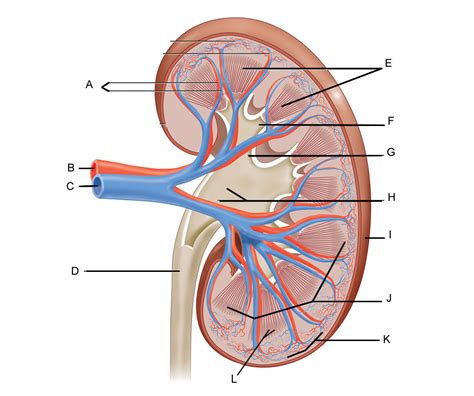
Table of Contents
Label the Diagram of the Kidney and Nephron Below: A Comprehensive Guide
Understanding the intricate workings of the human kidney and nephron is crucial for grasping the complexities of the urinary system and overall human physiology. This article provides a detailed, illustrated guide to labeling diagrams of both the kidney and the nephron, incorporating key anatomical structures and their functions. We'll also explore the physiological processes occurring within these structures, providing a holistic understanding of renal function. This guide is designed to be comprehensive, suitable for students, healthcare professionals, and anyone interested in learning more about this vital organ system.
The Kidney: Structure and Function
The kidney, a bean-shaped organ approximately the size of a fist, is the primary functional unit of the urinary system. Its primary roles include:
- Filtration: Removing waste products and excess water from the blood.
- Reabsorption: Returning essential nutrients, electrolytes, and water back into the bloodstream.
- Secretion: Actively transporting specific substances from the blood into the urine for excretion.
- Hormone Production: Producing hormones like erythropoietin (stimulates red blood cell production) and renin (regulates blood pressure).
Let's examine the key anatomical structures of the kidney, illustrated in the diagram below (insert a labeled diagram of the kidney here, including the following structures):
Key Structures of the Kidney:
- Renal Capsule: The tough, fibrous outer layer protecting the kidney.
- Renal Cortex: The outer region of the kidney, containing the glomeruli and convoluted tubules. This area is rich in blood vessels.
- Renal Medulla: The inner region of the kidney, composed of renal pyramids containing the loops of Henle and collecting ducts.
- Renal Pyramids: Triangular-shaped structures within the medulla, containing the collecting ducts.
- Renal Pelvis: A funnel-shaped structure that collects urine from the renal pyramids.
- Ureter: A tube that carries urine from the renal pelvis to the urinary bladder.
- Renal Artery: Brings oxygenated blood to the kidney.
- Renal Vein: Carries deoxygenated blood away from the kidney.
- Minor Calyces: Small cup-like structures that collect urine from the renal papillae.
- Major Calyces: Larger structures formed by the fusion of minor calyces.
- Renal Papillae: The apex of the renal pyramids where urine is drained into the minor calyces.
The Nephron: The Functional Unit of the Kidney
The nephron is the microscopic functional unit of the kidney. Millions of nephrons work together to perform the kidney's vital functions. Each nephron consists of two main parts:
Key Structures of the Nephron:
-
Renal Corpuscle (Malpighian Body): This is the initial filtering unit.
- Glomerulus: A network of capillaries where blood filtration occurs. The high pressure within the glomerulus forces fluids and small molecules out of the blood and into the Bowman's capsule.
- Bowman's Capsule (Glomerular Capsule): A cup-shaped structure surrounding the glomerulus, collecting the filtrate.
-
Renal Tubule: The filtrate passes through the renal tubule, where reabsorption and secretion occur.
- Proximal Convoluted Tubule (PCT): The first part of the renal tubule, where most reabsorption of water, glucose, amino acids, and other essential nutrients takes place.
- Loop of Henle (Nephron Loop): A U-shaped structure extending into the renal medulla. It plays a crucial role in concentrating the urine through countercurrent multiplication. This involves the establishment of an osmotic gradient within the medulla, allowing for efficient water reabsorption.
- Descending Limb of the Loop of Henle: Permeable to water but not to solutes. Water is reabsorbed passively due to the high osmolarity of the medullary interstitium.
- Ascending Limb of the Loop of Henle: Impermeable to water, but actively transports sodium, potassium, and chloride ions out of the tubule, contributing to the medullary osmotic gradient.
- Distal Convoluted Tubule (DCT): The final part of the renal tubule. Fine-tuning of electrolyte balance occurs here, regulated by hormones such as aldosterone.
- Collecting Duct: Receives filtrate from multiple nephrons and conducts it to the renal pelvis. The permeability of the collecting duct to water is regulated by antidiuretic hormone (ADH), affecting the final concentration of urine.
(Insert a labeled diagram of the nephron here, including all the structures listed above.)
Processes within the Nephron: Filtration, Reabsorption, and Secretion
The nephron performs three main processes:
1. Glomerular Filtration:
This process occurs in the renal corpuscle. Blood pressure forces water, small solutes (glucose, amino acids, ions), and waste products (urea, creatinine) from the glomerular capillaries into Bowman's capsule. Larger molecules like proteins and blood cells are generally excluded. The filtrate formed here is similar to blood plasma but without the large proteins.
2. Tubular Reabsorption:
As the filtrate flows through the renal tubule, essential substances are reabsorbed back into the bloodstream. This process is highly selective and involves both passive and active transport mechanisms. The proximal convoluted tubule is the primary site for reabsorption of water, glucose, amino acids, and ions. The loop of Henle plays a crucial role in reabsorbing water and establishing the medullary concentration gradient. The distal convoluted tubule fine-tunes the reabsorption of ions.
3. Tubular Secretion:
This process involves the active transport of certain substances from the peritubular capillaries into the renal tubule. This mechanism helps to remove additional waste products, such as hydrogen ions (H+), potassium ions (K+), and certain drugs from the blood, further refining the filtrate.
Hormonal Regulation of Renal Function
Several hormones play a vital role in regulating kidney function:
- Antidiuretic Hormone (ADH): Increases the permeability of the collecting duct to water, leading to increased water reabsorption and concentrated urine.
- Aldosterone: Promotes sodium reabsorption and potassium secretion in the distal convoluted tubule and collecting duct, influencing blood pressure and electrolyte balance.
- Renin-Angiotensin-Aldosterone System (RAAS): A complex hormonal system that regulates blood pressure. Renin, released by the kidneys in response to low blood pressure, triggers a cascade of events leading to aldosterone release and increased blood pressure.
- Parathyroid Hormone (PTH): Increases calcium reabsorption in the renal tubules.
- Atrial Natriuretic Peptide (ANP): Increases sodium excretion, promoting fluid loss and lowering blood pressure.
Clinical Significance
Understanding the structure and function of the kidney and nephron is essential for diagnosing and managing various renal diseases, including:
- Acute Kidney Injury (AKI): Sudden loss of kidney function.
- Chronic Kidney Disease (CKD): Gradual loss of kidney function over time.
- Glomerulonephritis: Inflammation of the glomeruli.
- Kidney Stones: Formation of crystals in the kidney.
- Urinary Tract Infections (UTIs): Infections affecting any part of the urinary tract.
Proper labeling of kidney and nephron diagrams is crucial for understanding the physiological processes involved in renal function and for comprehending the pathology of various kidney disorders. This knowledge is essential for healthcare professionals and students alike. Continued study and practice in identifying and labeling these structures will solidify your understanding of this critical organ system. Remember to consult reliable anatomical resources and utilize interactive diagrams for enhanced learning.
Latest Posts
Latest Posts
-
A Velocity Time Graph Shows How Velocity Changes Over
Apr 09, 2025
-
What Is Lambda In Physics Electric Field
Apr 09, 2025
-
Of What Value Is A Simple Stain
Apr 09, 2025
-
Which Structure Is Highlighted Nucleus Of Cardiac Muscle Fiber
Apr 09, 2025
-
Is Electric Conductor Acid Or Base
Apr 09, 2025
Related Post
Thank you for visiting our website which covers about Label The Diagram Of The Kidney And Nephron Below . We hope the information provided has been useful to you. Feel free to contact us if you have any questions or need further assistance. See you next time and don't miss to bookmark.