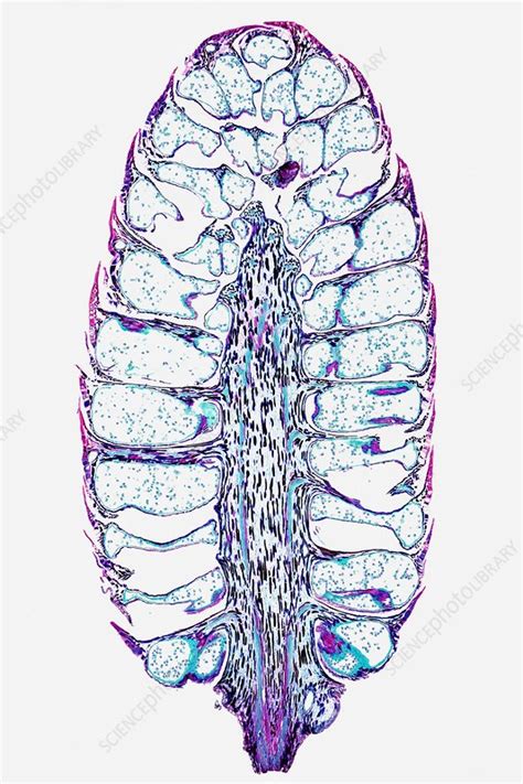
Male Pine Cone Under Microscope Labeled
Muz Play
Apr 05, 2025 · 6 min read

Table of Contents
Male Pine Cone Under the Microscope: A Detailed Exploration
The seemingly humble male pine cone, often overlooked in favor of its larger, more conspicuous female counterpart, reveals a world of intricate botanical beauty when viewed under a microscope. This article delves into the fascinating microscopic anatomy of the male pine cone, exploring its structures, functions, and significance in the reproductive cycle of pine trees. We'll cover everything from the individual pollen grains to the overall organization of the cone's microsporangia, providing a comprehensive understanding of this often-unseen component of the pine life cycle.
The Male Pine Cone: An Overview
Before diving into the microscopic details, let's establish a foundational understanding of the male pine cone itself. Unlike the woody, persistent female cones that bear seeds, male cones are typically smaller, softer, and ephemeral. They are responsible for producing and releasing vast quantities of pollen, the male gametophyte in the pine life cycle. These cones are usually found clustered together at the base of new shoots, often appearing in large numbers on the tree. Their delicate structure and temporary nature reflect their singular purpose: pollen production and dissemination. Their short lifespan ensures that pollen release occurs at the optimal time for fertilization.
Microscopic Anatomy of the Male Pine Cone: A Deep Dive
The true beauty of the male pine cone lies in its intricate microscopic structure. When observed under magnification, the arrangement of microsporangia, pollen sacs, and individual pollen grains becomes breathtakingly clear. Here's a detailed breakdown of what we would see under the microscope:
1. The Microsporangia: The Pollen Factories
The male cone's primary function is pollen production, and this takes place within specialized structures called microsporangia. These are elongated sacs, typically clustered together in groups within the cone's scales. Under a microscope, the microsporangia appear as distinct compartments filled with a mass of developing pollen grains. Their walls are relatively thin, allowing for the easy release of mature pollen. The arrangement and density of these microsporangia vary between pine species, adding to the diversity of the male cone's microscopic appearance. Their size and shape are crucial characteristics used in the taxonomic identification of different pine species.
2. Pollen Grains: The Male Gametophytes
The microsporangia are packed with pollen grains, the microscopic male gametophytes. These are not just simple cells; each pollen grain is a complex structure containing the genetic material necessary for fertilization. Under high magnification, the intricate surface details of the pollen grains become apparent. Many pine pollen grains exhibit characteristic air sacs, or bladders, which aid in wind dispersal. These bladders significantly increase the surface area, making the pollen grain lighter and allowing it to be carried further by air currents. The size and shape of these air sacs, along with the overall morphology of the pollen grain, are valuable taxonomic characters. The surface of the pollen grain itself can display a variety of patterns and textures, further aiding in species identification.
3. The Microsporophylls: The Supporting Structure
The microsporangia are borne on specialized leaf-like structures called microsporophylls. These are modified leaves arranged spirally on the central axis of the male cone. Under the microscope, the microsporophylls show clear differentiation from other vegetative leaves, with specialized tissues supporting the microsporangia. The arrangement and morphology of the microsporophylls contribute to the overall structure and appearance of the male cone. Their size and shape, along with the arrangement of the microsporangia on them, vary greatly between different pine species, providing useful clues for identification purposes. Analyzing these structures microscopically allows for a detailed examination of the developmental process and the overall organization of the male cone.
4. Tapetum: Nourishing the Pollen
Within the microsporangium, a specialized nutritive layer known as the tapetum plays a crucial role in pollen development. The tapetum cells provide nutrients and essential molecules to the developing pollen grains. Under the microscope, the tapetum is visible as a layer of cells surrounding the pollen mother cells (PMC). The tapetum undergoes various developmental changes and contributes to the formation of the pollen wall. Its structure and function are crucial for successful pollen production.
Microscopic Techniques for Studying Male Pine Cones
Several microscopic techniques are employed to study the intricacies of the male pine cone.
-
Light Microscopy: This is the most common method, providing high-resolution images of the overall structure of the cone, microsporangia, pollen grains, and microsporophylls. Different staining techniques can be used to enhance contrast and highlight specific cellular components.
-
Scanning Electron Microscopy (SEM): SEM provides incredibly detailed images of the surface textures of pollen grains and the microsporophylls, revealing fine details like the sculpturing on the pollen surface or the arrangement of stomata.
-
Transmission Electron Microscopy (TEM): TEM offers the highest magnification, allowing visualization of internal cell structures within pollen grains, providing insights into the details of the cell's organelles and their function in pollen development.
The Significance of Microscopic Analysis
Microscopic examination of male pine cones is crucial for several reasons:
-
Taxonomy and Identification: The microscopic characteristics of pollen grains and other structures are valuable tools for identifying and classifying different pine species. Slight variations in pollen morphology can distinguish between closely related species.
-
Pollination Biology: Studying pollen morphology helps in understanding the mechanisms of pollination in pines. The presence of air sacs, for example, indicates wind pollination, while other features may indicate adaptations to different pollinating agents.
-
Evolutionary Studies: Comparing the microscopic structure of male cones across different pine species can provide insights into their evolutionary relationships and the diversification of pines over time. Microscopic analysis of fossils can shed light on the evolution of this important reproductive structure.
-
Paleobotany: Fossil pollen grains (palynomorphs) are important sources of information about past vegetation and climate. Microscopic examination is essential in identifying fossil pollen, allowing for reconstructions of past ecosystems.
Conclusion: Unveiling the Hidden World
The male pine cone, while often overlooked, reveals a stunning level of complexity under the microscope. From the intricate organization of the microsporangia to the detailed architecture of the individual pollen grains, a microscopic examination unveils a world of botanical wonder. This detailed exploration is crucial not only for understanding the reproductive biology of pine trees but also for various applications in taxonomy, evolutionary biology, and paleobotany. The next time you encounter a seemingly simple male pine cone, remember the hidden complexities waiting to be uncovered with the aid of a microscope. The seemingly simple structure is a testament to the intricate design found within the natural world. Further research and microscopic studies are continuously revealing new insights into the secrets held within these tiny, yet significant, reproductive structures.
Latest Posts
Latest Posts
-
Find The Equation Of The Tangent Plane To The Surface
Apr 05, 2025
-
Third Trophic Level In The Food Chain
Apr 05, 2025
-
How To Solve 3 Equations 3 Unknowns
Apr 05, 2025
-
How Many Times More Acidic Is Ph3 Than Ph5
Apr 05, 2025
-
What Instruments Are Used To Measure Humidity
Apr 05, 2025
Related Post
Thank you for visiting our website which covers about Male Pine Cone Under Microscope Labeled . We hope the information provided has been useful to you. Feel free to contact us if you have any questions or need further assistance. See you next time and don't miss to bookmark.
