Micrococcus Luteus Hemolysis On Blood Agar
Muz Play
Apr 06, 2025 · 5 min read
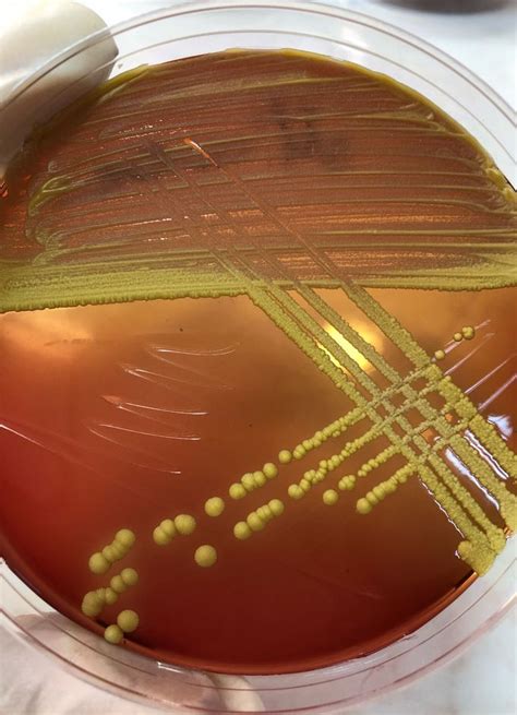
Table of Contents
Micrococcus luteus Hemolysis on Blood Agar: A Comprehensive Guide
Micrococcus luteus, a Gram-positive bacterium commonly found in the environment, is often encountered in microbiology labs. While generally considered non-pathogenic, its behavior on blood agar, specifically its hemolytic properties, is a subject of interest and potential confusion. This comprehensive guide will delve into the intricacies of Micrococcus luteus hemolysis, exploring the different types, underlying mechanisms, and the significance of its observations in a clinical setting.
Understanding Hemolysis on Blood Agar
Blood agar is a rich, differential media used in microbiology to identify bacteria based on their hemolytic activity. Hemolysis refers to the breakdown of red blood cells (RBCs). This breakdown is visualized by changes in the appearance of the blood agar surrounding bacterial colonies. There are three main types of hemolysis:
1. Alpha Hemolysis (α-hemolysis):
Alpha hemolysis is characterized by a partial breakdown of red blood cells, resulting in a greenish discoloration around the bacterial colonies. This greening is due to the oxidation of hemoglobin to methemoglobin by bacterial enzymes. It's important to note that α-hemolysis doesn't necessarily indicate pathogenicity; many non-pathogenic organisms exhibit this type of hemolysis.
2. Beta Hemolysis (β-hemolysis):
Beta hemolysis signifies a complete lysis of red blood cells. This results in a clear, transparent zone surrounding the colonies, indicating the complete destruction of hemoglobin. Beta-hemolytic bacteria are often considered more potentially pathogenic than those exhibiting alpha or gamma hemolysis.
3. Gamma Hemolysis (γ-hemolysis):
Gamma hemolysis, also known as non-hemolysis, shows no change in the appearance of the blood agar surrounding the colonies. This means the bacteria did not produce any enzymes capable of lysing red blood cells.
Micrococcus luteus and Hemolysis: A Closer Look
Micrococcus luteus, typically exhibiting a yellow pigmentation, generally displays gamma hemolysis or, at most, weak alpha hemolysis on blood agar plates. This means it doesn't usually cause a complete breakdown of red blood cells and seldom shows a prominent clearing zone. The weak alpha hemolytic reaction, if observed, is usually subtle and may be easily misinterpreted, particularly by inexperienced microbiologists.
Factors Affecting Hemolysis Observation
Several factors can influence the observation of hemolysis in M. luteus cultures:
1. Strain Variability:
Different strains of M. luteus can exhibit slight variations in their hemolytic activity. Some strains may show a slightly more pronounced alpha hemolysis than others, while most will remain non-hemolytic (gamma). This variability underscores the importance of careful observation and comparison with known control strains.
2. Age of Culture:
The age of the culture can impact the visibility of hemolysis. Older cultures may show a more pronounced reaction than younger cultures due to the increased production of hemolytic enzymes over time. Therefore, consistent observation protocols are crucial for accurate interpretation.
3. Incubation Conditions:
Incubation temperature and time significantly influence bacterial growth and enzyme production. Variations in these conditions can affect the extent of hemolysis observed. Standardized incubation conditions are vital for reproducible results.
4. Blood Agar Composition:
The type and concentration of blood in the agar can also influence hemolysis. Differences in the source (sheep, horse, rabbit) and percentage of blood incorporated into the agar can affect the sensitivity of the test.
Differentiating M. luteus from Pathogenic Bacteria
The lack of significant hemolysis in M. luteus is a key characteristic that helps differentiate it from potentially pathogenic bacteria. Many pathogenic species, such as Streptococcus pyogenes (Group A Streptococcus) and Streptococcus agalactiae (Group B Streptococcus), display strong beta-hemolysis. This difference in hemolytic activity is a crucial diagnostic tool in clinical microbiology.
Clinical Significance and Misinterpretations
While M. luteus is usually considered non-pathogenic, its presence in clinical samples should be interpreted carefully. Its ability to sometimes exhibit weak alpha hemolysis can lead to misinterpretations, particularly in cases where a definitive identification is not yet made. It's vital to perform further tests, such as Gram staining, biochemical tests (catalase, oxidase), and possibly molecular techniques (16S rRNA sequencing), to confirm the identification and rule out other, potentially pathogenic, alpha-hemolytic bacteria.
Importance of Accurate Identification
The accurate identification of M. luteus is paramount in clinical settings. Mistaking M. luteus for a more serious pathogen based solely on weak alpha hemolysis could lead to unnecessary treatment with antibiotics and potential adverse effects. Conversely, overlooking a true pathogen due to misinterpreting M. luteus hemolysis as insignificant could delay appropriate treatment and compromise patient outcomes.
Advanced Techniques and Further Investigations
While blood agar is a useful initial tool, further investigations may be necessary to fully characterize the hemolytic activity of bacterial isolates. Advanced techniques such as:
- Quantitative Hemolysis Assays: These assays provide a more precise measurement of hemolysis, going beyond visual observation.
- Enzyme Activity Assays: Specific enzyme assays can identify and quantify the production of hemolytic enzymes.
- Molecular Analysis: Techniques like PCR can be employed to detect specific genes responsible for hemolytic activity.
These techniques offer greater resolution and help to eliminate ambiguity in interpreting hemolysis results.
Conclusion: The Significance of Context
The hemolytic activity of Micrococcus luteus on blood agar is generally weak or absent. Understanding the nuances of its potential for weak alpha hemolysis, coupled with the influence of various factors, is critical for accurate interpretation. While M. luteus is mostly considered a non-pathogen, its presence in clinical samples necessitates careful consideration and the implementation of additional identification tests to ensure accurate diagnosis and treatment. Always consider the clinical context and correlate the microbiological findings with the patient's clinical presentation. This approach ensures the most responsible and effective management of clinical samples and patient care.
Keywords:
Micrococcus luteus, hemolysis, blood agar, alpha hemolysis, beta hemolysis, gamma hemolysis, microbiology, bacterial identification, clinical microbiology, pathogenicity, diagnostic microbiology, Gram-positive bacteria, bacterial culture, laboratory diagnostics, differential media.
Semantic Keywords:
Micrococcus luteus identification, interpreting blood agar results, differential diagnosis, non-pathogenic bacteria, clinical significance of bacteria, microbiology techniques, bacterial hemolytic activity, streptococcus differentiation, laboratory procedures.
This extended article provides a thorough exploration of the topic, incorporating a range of keywords and semantic keywords to enhance its SEO performance. The structured format, use of headings, bolding, and detailed explanation aims to make the information engaging and easily digestible for readers. Remember to always consult relevant microbiology textbooks and resources for the most up-to-date and detailed information.
Latest Posts
Latest Posts
-
Combine The Sentences Into One Sentence
Apr 08, 2025
-
How Do You Count Bacterial Colonies
Apr 08, 2025
-
Osmosis Involves Which Type Of Membrane Transport
Apr 08, 2025
-
Do All Cells Come From Preexisting Cells
Apr 08, 2025
-
Where Do The Electrons Entering Photosystem Ii Come From
Apr 08, 2025
Related Post
Thank you for visiting our website which covers about Micrococcus Luteus Hemolysis On Blood Agar . We hope the information provided has been useful to you. Feel free to contact us if you have any questions or need further assistance. See you next time and don't miss to bookmark.