The Fourth Ventricle Is Represented By Letter
Muz Play
Mar 24, 2025 · 6 min read
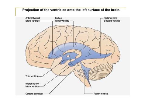
Table of Contents
The Fourth Ventricle: Anatomy, Function, and Clinical Significance Represented by the Letter 'V'
The fourth ventricle, a crucial component of the central nervous system, is often represented by the letter 'V' in anatomical diagrams and discussions. This article delves into the detailed anatomy, physiology, and clinical significance of this vital structure, emphasizing its representation and importance within the broader context of the brain's ventricular system.
H2: Understanding the Ventricular System: The 'V' of the Fourth Ventricle
The brain's ventricular system is a network of interconnected cavities filled with cerebrospinal fluid (CSF). This system plays a critical role in protecting the brain, providing buoyancy, and transporting nutrients and waste products. The system comprises four ventricles: two lateral ventricles, the third ventricle, and the fourth ventricle. Our focus is on the fourth ventricle, often visualized and symbolically represented by 'V' due to its diamond-like shape in many anatomical cross-sections. The 'V' serves as a helpful mnemonic device for remembering its location and overall form within the complex architecture of the brainstem.
H2: Anatomy of the Fourth Ventricle: A Detailed Exploration of the 'V'
The fourth ventricle is located within the brainstem, specifically between the pons and the medulla oblongata dorsally, and the cerebellum ventrally. Its shape is roughly rhomboid, resembling a 'V' when viewed in certain anatomical planes. The floor of the fourth ventricle is formed by the rhomboid fossa, a part of the brainstem floor. Key anatomical features contributing to the 'V' shape include:
H3: The Rhomboid Fossa: Forming the Floor of the 'V'
The rhomboid fossa, the floor of the fourth ventricle, is a significant landmark. Its features, including the median sulcus, facial colliculus, and hypoglossal trigone, all contribute to its overall shape and are crucial for understanding the neurovascular relationships within the ventricle.
H3: Cerebellar Peduncles: Contributing to the 'V' Structure
The superior, middle, and inferior cerebellar peduncles, tracts of white matter connecting the cerebellum to the brainstem, contribute to the boundaries of the fourth ventricle, influencing the overall 'V' shape. Understanding their course is essential for understanding the circulation of CSF and the potential for blockage in clinical conditions.
H3: Apertures of the Fourth Ventricle: The CSF Pathway
The fourth ventricle communicates with the subarachnoid space through three apertures: the median aperture (foramen of Magendie) and two lateral apertures (foramina of Luschka). These apertures are vital for the flow of CSF from the ventricular system into the subarachnoid space, surrounding the brain and spinal cord. Blockages in these apertures can lead to hydrocephalus, a potentially life-threatening condition. These apertures are integral parts of the 'V' structure, defining its borders and functional connections.
H2: Function of the Fourth Ventricle: Beyond the 'V'
While its shape—often represented by the letter 'V'—is a helpful anatomical marker, the fourth ventricle's primary function centers around cerebrospinal fluid (CSF) circulation and its role in maintaining homeostasis within the central nervous system.
H3: Cerebrospinal Fluid (CSF) Production and Circulation
The fourth ventricle receives CSF from the third ventricle via the cerebral aqueduct. Within the fourth ventricle, the CSF is further circulated and then exits through the median and lateral apertures to enter the subarachnoid space. The continuous production and circulation of CSF are essential for brain cushioning, nutrient transport, waste removal, and maintaining a stable intracranial pressure. The 'V' shape, therefore, indirectly facilitates this crucial process.
H3: Neurotransmitter Production and Regulation
The floor of the fourth ventricle contains numerous nuclei of cranial nerves, playing roles in vital bodily functions including breathing, heart rate, and swallowing. This suggests a possible role for the fourth ventricle in the production and regulation of neurotransmitters influencing these critical functions. The 'V' shape simply houses this complex functionality.
H2: Clinical Significance: When the 'V' is Compromised
Various pathological conditions can affect the fourth ventricle, impacting CSF flow and potentially leading to severe neurological consequences.
H3: Hydrocephalus: Blockage and Impaired CSF Flow
Hydrocephalus, characterized by an excess of CSF within the ventricular system, can result from blockages at the apertures of the fourth ventricle. These blockages prevent proper CSF drainage into the subarachnoid space, causing the ventricles to expand, potentially leading to increased intracranial pressure and severe neurological damage. The 'V' shape, in this instance, highlights the critical location of the outflow points crucial for maintaining proper CSF dynamics.
H3: Tumors and Cysts: Space-Occupying Lesions
Tumors and cysts arising within or near the fourth ventricle can obstruct CSF flow, leading to hydrocephalus. These lesions can compress surrounding brain structures, resulting in neurological deficits depending on their location and size. The 'V' region's proximity to vital brainstem structures underscores the severity of compression from lesions in this area.
H3: Dandy-Walker Malformation: Congenital Anomaly
Dandy-Walker malformation, a congenital anomaly, involves abnormal development of the fourth ventricle and cerebellum. This malformation can lead to hydrocephalus and neurological deficits, highlighting the crucial role of the fourth ventricle in normal brain development. The 'V' shape in this case is dramatically altered, reflecting the developmental defect.
H2: Imaging Techniques and the 'V': Visualization of the Fourth Ventricle
Several neuroimaging techniques allow for detailed visualization of the fourth ventricle, confirming its anatomical features and aiding in the diagnosis of pathological conditions.
H3: Magnetic Resonance Imaging (MRI): A High-Resolution View of the 'V'
MRI provides excellent soft tissue contrast, allowing detailed visualization of the fourth ventricle's structure, including its floor (rhomboid fossa), boundaries, and apertures. This technique is crucial in identifying lesions and assessing the patency of CSF pathways. The 'V' can be clearly identified in various MRI sequences.
H3: Computed Tomography (CT): Assessing Density Changes
CT scanning, while providing less detail than MRI, is valuable in identifying calcifications, hemorrhages, and other density changes within or around the fourth ventricle. This technique can be used as a rapid screening tool, potentially highlighting the need for more detailed investigations like MRI. Even the 'V' shape can be suggested on CT, especially in cases of abnormality.
H2: The Future of Fourth Ventricle Research: Expanding our Understanding of the 'V'
Ongoing research continues to unravel the intricate complexities of the fourth ventricle's function and its role in various neurological processes. Advances in neuroimaging techniques and molecular biology are promising to provide a deeper understanding of this vital structure and its contribution to brain health.
H3: Advanced Neuroimaging Techniques
Future advancements in neuroimaging, such as diffusion tensor imaging (DTI) and functional MRI (fMRI), promise to provide even more detailed information about the connectivity and functional roles of the fourth ventricle and surrounding structures. The 'V' shape will be further clarified through these improved visualizations.
H3: Molecular and Genetic Studies
Molecular and genetic studies are crucial for understanding the developmental processes that lead to malformations of the fourth ventricle and other related conditions, leading to improved diagnostic and therapeutic strategies. Research on the 'V' area might uncover genetic factors contributing to its formation and function.
H2: Conclusion: The Enduring Significance of the 'V'
The fourth ventricle, often represented by the letter 'V' due to its shape, is a crucial structure within the central nervous system. Its role in cerebrospinal fluid circulation and its proximity to vital brainstem structures make it a critical component of neurological function. Understanding its anatomy, physiology, and clinical significance is vital for clinicians involved in the diagnosis and treatment of neurological conditions. The 'V' serves as a valuable mnemonic and visual aid in navigating the complexities of this essential structure. Further research will surely continue to illuminate the many secrets hidden within this seemingly simple, ‘V’ shaped chamber.
Latest Posts
Related Post
Thank you for visiting our website which covers about The Fourth Ventricle Is Represented By Letter . We hope the information provided has been useful to you. Feel free to contact us if you have any questions or need further assistance. See you next time and don't miss to bookmark.
