The Thoracic Cavity Is A Subdivision Of The
Muz Play
Mar 18, 2025 · 6 min read
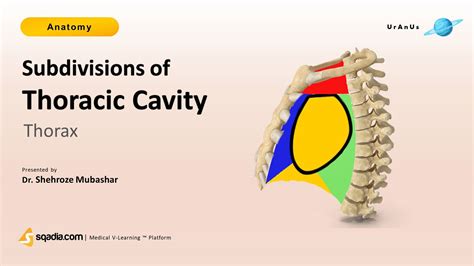
Table of Contents
The Thoracic Cavity: A Subdivision of the Ventral Body Cavity
The human body is a marvel of intricate organization, with various cavities housing vital organs and systems. Understanding the arrangement of these cavities is crucial for comprehending anatomy, physiology, and pathology. This article delves deep into the thoracic cavity, exploring its location, boundaries, contents, and significance as a crucial subdivision of the larger ventral body cavity.
The Ventral Body Cavity: A Broad Overview
Before focusing on the thoracic cavity, let's establish its broader context within the ventral body cavity. The ventral body cavity, also known as the coelom, is a large, fluid-filled space that occupies the anterior aspect of the body. It's fundamentally divided into two main cavities:
- Thoracic Cavity: The superior compartment, containing the heart, lungs, and associated structures.
- Abdominopelvic Cavity: The inferior compartment, encompassing the abdominal and pelvic cavities. The abdominal cavity houses the digestive organs, while the pelvic cavity contains the urinary bladder, reproductive organs, and rectum.
These cavities are not completely separate; they are continuous with each other. However, the diaphragm, a dome-shaped muscle, forms a significant boundary separating the thoracic and abdominopelvic cavities.
Defining the Thoracic Cavity: Boundaries and Divisions
The thoracic cavity, also called the chest cavity or thorax, is a cone-shaped space enclosed by the ribs, sternum, vertebral column, and diaphragm. Its boundaries are precisely defined:
- Superiorly: The thoracic inlet, formed by the first rib, manubrium of the sternum, and the first thoracic vertebra.
- Inferiorly: The diaphragm, a crucial muscle responsible for breathing. Its dome shape presses up against the lungs and other thoracic organs.
- Anteriorly: The sternum and costal cartilages (connecting the ribs to the sternum).
- Posteriorly: The thoracic vertebrae.
- Laterally: The ribs and intercostal muscles (muscles between the ribs).
Within the thoracic cavity, three smaller compartments are identified:
- Mediastinum: The central compartment, a relatively thick partition of tissues and organs located between the lungs. It houses the heart, great vessels (aorta, vena cavae, pulmonary arteries and veins), trachea, esophagus, thymus, and lymph nodes.
- Right Pleural Cavity: The space surrounding the right lung, lined by the pleural membrane.
- Left Pleural Cavity: The space surrounding the left lung, similarly lined by the pleural membrane.
The Pleura: A Protective Lining
The pleural cavities are not simply empty spaces; they are lined by a serous membrane called the pleura. This membrane consists of two layers:
- Parietal Pleura: The outer layer, adhering to the thoracic wall, diaphragm, and mediastinum.
- Visceral Pleura: The inner layer, intimately covering the surface of each lung.
Between these two layers is a potential space, the pleural space, containing a small amount of serous fluid. This fluid acts as a lubricant, reducing friction during lung expansion and contraction. The integrity of the pleural membranes is essential for proper lung function. Damage to the pleura, such as in pneumothorax (collapsed lung), can significantly impair breathing.
The Mediastinum: The Heart's Central Domain
The mediastinum, the central compartment of the thoracic cavity, holds a multitude of vital structures. Its location and contents are critical to understanding cardiovascular, respiratory, and lymphatic systems. The mediastinum is further subdivided into superior and inferior mediastina:
- Superior Mediastinum: This region extends from the thoracic inlet to the sternal angle. It contains the superior vena cava, aortic arch, brachiocephalic vessels, trachea, esophagus, thymus, and phrenic and vagus nerves.
- Inferior Mediastinum: Situated below the sternal angle, it's further divided into anterior, middle, and posterior compartments. The anterior mediastinum is largely filled with connective tissue. The middle mediastinum is the location of the heart, pericardium (the protective sac surrounding the heart), and great vessels entering and leaving the heart. The posterior mediastinum houses the descending aorta, esophagus, and thoracic duct.
The Pericardium: Protecting the Heart
The heart, a crucial pump driving blood circulation, is enclosed within a tough, fibrous sac called the pericardium. This sac provides protection and helps prevent overfilling of the heart. The pericardium is composed of two main layers:
- Fibrous Pericardium: The outermost, tough, protective layer.
- Serous Pericardium: A thinner, more delicate layer with two sublayers: the parietal pericardium (lining the fibrous pericardium) and the visceral pericardium (epicardium, adhering to the heart's surface). Between these layers lies the pericardial cavity, containing a small amount of fluid that lubricates the heart's movement.
Respiratory System Within the Thoracic Cavity
The lungs, the primary organs of respiration, occupy the majority of the space within the thoracic cavity. Each lung is encased in its own pleural cavity and is further divided into lobes: the right lung has three lobes, while the left lung has two. The lungs are responsible for gas exchange, taking in oxygen and releasing carbon dioxide. Their structure, with millions of alveoli (tiny air sacs), provides a vast surface area for efficient gas exchange. The trachea, branching into two main bronchi, conducts air to and from the lungs.
Cardiovascular System in the Thoracic Cavity
The heart, located within the mediastinum, is the central organ of the cardiovascular system. Its location within the protective pericardium is crucial to its function. Major blood vessels, including the aorta (carrying oxygenated blood away from the heart), vena cavae (returning deoxygenated blood to the heart), and pulmonary arteries and veins (involved in pulmonary circulation), are also found within the thoracic cavity. These vessels form a complex network delivering oxygen and nutrients to the body's tissues and removing waste products.
Neurological Structures in the Thoracic Cavity
Several crucial nerves traverse the thoracic cavity, providing innervation to various organs and tissues. The vagus nerve, for example, plays a significant role in parasympathetic control of heart rate and digestive function. The phrenic nerves innervate the diaphragm, vital for respiration. The sympathetic chain ganglia, part of the sympathetic nervous system, also lie within the thoracic cavity, influencing cardiovascular and respiratory responses.
Lymphatic System in the Thoracic Cavity
The thoracic cavity houses elements of the lymphatic system, which plays a critical role in immunity. The thymus, a gland vital for T-cell maturation, resides in the superior mediastinum during childhood and adolescence. Lymph nodes are scattered throughout the mediastinum and pleural cavities, filtering lymph and helping combat infections. The thoracic duct, the largest lymphatic vessel, collects lymph from the lower body and empties it into the venous system near the heart.
Clinical Significance of the Thoracic Cavity
Understanding the thoracic cavity's anatomy and contents is crucial in various medical contexts. Conditions affecting this region, such as:
- Pneumonia: Infection of the lung tissue.
- Pleurisy: Inflammation of the pleura.
- Pneumothorax: Collapsed lung due to air in the pleural cavity.
- Cardiac Tamponade: Accumulation of fluid in the pericardial cavity, compressing the heart.
- Lung Cancer: Malignant growth in the lung tissue.
- Aortic Aneurysm: Bulging or weakening of the aorta.
require a thorough understanding of the anatomical relationships within the thoracic cavity for accurate diagnosis and treatment. Imaging techniques like chest X-rays and CT scans are frequently employed to visualize structures within the thoracic cavity and aid in diagnosis.
Conclusion: A Complex and Vital Space
The thoracic cavity, a vital subdivision of the ventral body cavity, is a complex and intricately organized space housing several essential organ systems. Its boundaries, contents, and relationships with neighboring cavities are crucial to understanding human anatomy and physiology. From the protective pleural membranes surrounding the lungs to the intricate arrangement of the mediastinum and its contents, including the heart and great vessels, the thoracic cavity's structure ensures the efficient functioning of crucial life processes. A deep comprehension of this anatomical region is fundamental to clinical practice and further scientific exploration of human biology. Future research may unravel even more about the nuanced interactions between the various structures within this vital cavity, improving diagnostics, treatments, and overall human health.
Latest Posts
Latest Posts
-
Work Done By An Electric Field
Mar 18, 2025
-
How Much Energy To Be At Zero Kinetic Energy
Mar 18, 2025
-
Current As A Function Of Time
Mar 18, 2025
-
Ode To Billy Joe Lyrics Meaning
Mar 18, 2025
-
Where Does The Light Independent Reaction Take Place
Mar 18, 2025
Related Post
Thank you for visiting our website which covers about The Thoracic Cavity Is A Subdivision Of The . We hope the information provided has been useful to you. Feel free to contact us if you have any questions or need further assistance. See you next time and don't miss to bookmark.
