Through Which Microscope Were Cells First Observed
Muz Play
Apr 05, 2025 · 6 min read

Table of Contents
Through Which Microscope Were Cells First Observed? A Deep Dive into the History of Cell Discovery
The discovery of the cell, the fundamental unit of life, is a pivotal moment in the history of biology. Understanding how this discovery came about requires exploring the evolution of microscopy and the brilliant minds who pushed the boundaries of scientific observation. While pinpointing the exact microscope used is impossible, we can definitively trace the journey to the first cell observations, highlighting the crucial role of specific microscope designs and advancements.
The Dawn of Microscopy: Early Instruments and Limitations
Before diving into the cell's discovery, we need to understand the state of microscopy in the 17th century. Early microscopes were simple, single-lens devices, often referred to as simple microscopes. These instruments, while rudimentary by modern standards, were revolutionary for their time. They allowed for magnification beyond what the naked eye could achieve, opening up a new world of microscopic detail. However, these early microscopes suffered from significant limitations:
- Chromatic aberration: This resulted in colored fringes around the observed object, blurring the image and hindering accurate observation.
- Spherical aberration: This caused distortion due to the curvature of the lens, further compromising the image quality.
- Limited magnification: Even the best simple microscopes of the time could only achieve relatively low magnification, limiting the detail observable.
Despite these limitations, these simple microscopes laid the groundwork for future advancements. Experimentation with lens design and construction gradually improved image quality and magnification, paving the way for the discovery of cells.
Robert Hooke and the "Micrographia": The First Glimpse of Cells
The year 1665 marks a significant turning point. Robert Hooke, a prominent English scientist, published his groundbreaking work, "Micrographia". This book detailed numerous observations made using his compound microscope, a significant advancement over the simple microscope. Hooke's compound microscope employed two lenses:
- Objective lens: Located closest to the specimen, this lens initially magnified the image.
- Eyepiece lens: This lens further magnified the image produced by the objective lens.
This two-lens system provided significantly higher magnification compared to simple microscopes, allowing Hooke to observe finer details. In "Micrographia," Hooke described observing thin slices of cork under his compound microscope. He noted that the cork's structure resembled a honeycomb, comprised of tiny compartments that he termed "cells" — a term derived from the Latin word "cellula," meaning "small room."
Hooke's Compound Microscope: A Closer Look
While the exact specifications of Hooke's microscope are not precisely known, it's understood that his design represented a considerable leap in microscope technology. Although plagued by the same limitations as simple microscopes (chromatic and spherical aberration), his compound microscope provided sufficient magnification to reveal the cellular structure of cork. This achievement is monumental, marking the first documented observation of cells. However, it's important to note that Hooke observed only the cell walls of dead plant cells; he didn't observe the living cellular contents.
Antonie van Leeuwenhoek: Observing Living Cells
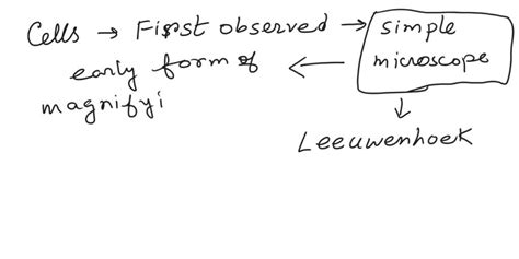
While Robert Hooke's work laid the foundation, it was Antonie van Leeuwenhoek, a Dutch draper and self-taught scientist, who took microscopy to a new level and observed living cells. Leeuwenhoek's microscopes were simple microscopes, but he possessed exceptional skill in crafting high-quality lenses with remarkable magnifying power. His lenses achieved significantly higher magnification than Hooke's compound microscope, although with a narrower field of view.
Leeuwenhoek meticulously documented his observations of various specimens, including pond water, rainwater, and even scrapings from his teeth. His detailed descriptions revealed a plethora of microscopic organisms, which he termed "animalcules." These "animalcules" were, in fact, living single-celled organisms, representing the first observations of living cells and providing crucial evidence for the existence of microorganisms.
Leeuwenhoek's Simple Microscopes: A Masterpiece of Craftsmanship
Leeuwenhoek's success stemmed from his exceptional lens-making skills. He developed techniques for grinding and polishing lenses that produced remarkable magnification, often exceeding 200x. His microscopes were simple in design, consisting of a single, powerful lens mounted in a small metal frame. This simplicity, combined with his meticulous observation techniques, allowed him to make groundbreaking discoveries, showcasing the potential of even simple microscopes in capable hands.
The Significance of Both Discoveries
Both Hooke and Leeuwenhoek contributed significantly to the discovery of the cell, each in their own way. Hooke's compound microscope revealed the cellular structure in dead plant material, providing the initial framework for understanding cellular organization. Leeuwenhoek's simple microscope, however, allowed for the observation of living cells, fundamentally changing our understanding of life's diversity and complexity. Neither microscope was perfect, but their limitations were overcome by the meticulous observation and scientific ingenuity of these pioneering scientists.
From Early Microscopes to Modern Microscopy
The microscopes used by Hooke and Leeuwenhoek were crude by today's standards. However, they were groundbreaking for their time. Subsequent advancements in lens design, including the development of achromatic lenses (which corrected chromatic aberration), and the invention of the electron microscope (offering vastly greater magnification), have revolutionized microscopy, allowing for much more detailed observations of cellular structures and processes. The advancements in microscopy continue to this day with technologies like super-resolution microscopy pushing the limits of resolution.
The Legacy of Cell Discovery: A Continuous Exploration
The discovery of the cell, through the efforts of Hooke and Leeuwenhoek using their respective microscopes, wasn't just a single event; it was the dawn of a new era in biological understanding. It established the cell theory, a cornerstone of modern biology, which postulates that:
- All living organisms are composed of one or more cells.
- The cell is the basic unit of structure and organization in organisms.
- Cells arise from pre-existing cells.
This understanding, stemming from the first observations through relatively simple microscopes, has had a profound impact on numerous fields, including medicine, genetics, and biotechnology. The ongoing development of microscopy continues to reveal new insights into the complex workings of cells, pushing the boundaries of scientific knowledge and influencing our comprehension of life itself.
Conclusion: Honoring the Pioneers
While we cannot definitively say which specific microscope was "the" microscope through which cells were first observed, the contributions of Robert Hooke and Antonie van Leeuwenhoek are undeniable. Hooke's compound microscope provided the first glimpse of the cellular structure, and Leeuwenhoek's simple microscope revealed the dynamic world of living cells. Their work, driven by curiosity and meticulous observation, laid the foundation for modern cell biology and continues to inspire generations of scientists. Their legacy serves as a testament to the power of observation, ingenuity, and the relentless pursuit of knowledge. The quest to understand the cell, initiated with relatively simple tools, has led to the sophisticated microscopy techniques we have today, underscoring the continuous evolution of scientific exploration.
Latest Posts
Latest Posts
-
Worksheet Writing And Balancing Chemical Reactions
Apr 06, 2025
-
Difference Between Bronsted Acid And Lewis Acid
Apr 06, 2025
-
Buffer Region Of A Titration Curve
Apr 06, 2025
-
Poe Was Considered The Father Of
Apr 06, 2025
-
Half Of A Yellow Sun Ugwu
Apr 06, 2025
Related Post
Thank you for visiting our website which covers about Through Which Microscope Were Cells First Observed . We hope the information provided has been useful to you. Feel free to contact us if you have any questions or need further assistance. See you next time and don't miss to bookmark.
