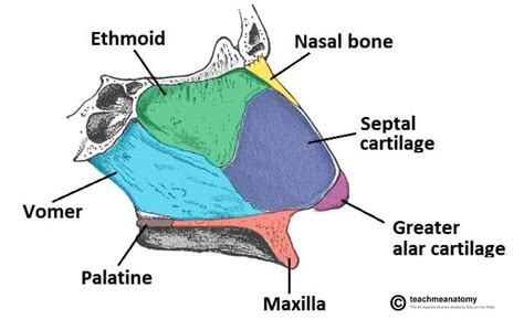
Two Bones That Form The Nasal Septum
Muz Play
Apr 04, 2025 · 6 min read

Table of Contents
Two Bones That Form the Nasal Septum: A Deep Dive into Anatomy, Function, and Clinical Significance
The nasal septum, that crucial midline structure dividing the nasal cavity into two symmetrical halves, is a complex anatomical feature vital for proper breathing and olfaction. While often perceived as a singular entity, the septum is actually formed by a fascinating interplay of several bony and cartilaginous components. This article focuses primarily on the two primary bony contributors: the vomer and the perpendicular plate of the ethmoid bone. We will delve into their individual anatomy, their combined contribution to the nasal septum, common deviations and disorders affecting this delicate structure, and the clinical implications thereof.
The Vomer: A Keystone of the Nasal Septum
The vomer, deriving its name from the Latin word for "plowshare," is a thin, roughly ploughshare-shaped bone located in the midline of the nasal cavity. It forms the posteroinferior portion of the nasal septum, contributing significantly to its structural integrity.
Anatomical Features of the Vomer:
- Shape and Size: The vomer's shape is far from uniform. It presents with a broad, superior portion which articulates with other bones, and narrows inferiorly, ending in a sharp point. Its size and exact shape can vary slightly between individuals.
- Articulations: The vomer's articulations are crucial to its role in forming the septum. It articulates superiorly with the sphenoid bone (specifically, the rostrum of the sphenoid), laterally with the medial pterygoid plates of the sphenoid, and inferiorly with the palatine bones and the maxillary bones. The articulation with the perpendicular plate of the ethmoid forms the critical junction of the bony septum.
- Surface Features: The vomer exhibits a slightly concave medial surface, facing the nasal cavity. Its lateral surfaces articulate with the aforementioned bones. It also possesses grooves and ridges that accommodate the blood vessels and nerves supplying the nasal mucosa.
- Development: Ossification of the vomer begins during fetal development and continues into postnatal life. This complex process involves multiple ossification centers, highlighting the intricate development of this seemingly simple bone.
The Perpendicular Plate of the Ethmoid Bone: The Superior Septum Foundation
The ethmoid bone, a complex structure situated anterior to the sphenoid bone, contributes significantly to the nasal septum via its perpendicular plate. This plate forms the superior and anterior portions of the bony septum, articulating with the vomer below and the nasal bones and cartilage anteriorly.
Anatomical Features of the Perpendicular Plate:
- Location and Shape: The perpendicular plate descends from the cribriform plate of the ethmoid bone (which houses olfactory foramina). It's a thin, flat plate of bone, roughly rectangular in shape, that forms the superior part of the nasal septum.
- Articulations: As mentioned, it articulates with the vomer inferiorly, creating the keystone junction of the bony septum. It also articulates with the nasal bones anteriorly, and with the crista galli (a superior projection of the ethmoid) superiorly. This complex interplay of articulations reinforces the stability of the septum.
- Relationship to the Olfactory System: Its proximity to the cribriform plate and its role in supporting the nasal cavity makes it crucial to the olfactory pathway. Damage to the perpendicular plate can potentially disrupt olfactory function.
- Development: Like the vomer, the perpendicular plate undergoes ossification during fetal and postnatal development. This is tightly regulated and any disruptions during development can lead to septal anomalies.
The Combined Role of the Vomer and Perpendicular Plate
The vomer and the perpendicular plate of the ethmoid bone work synergistically to create the primary bony framework of the nasal septum. The perpendicular plate forms the superior aspect, while the vomer forms the inferior aspect. The junction between these two bones is critical and any deviation or malformation here can have significant clinical implications. The cartilaginous septum, the septal cartilage, then attaches to this bony framework, completing the septum's structure.
Clinical Significance of the Bony Septum:
- Septal Deviation: One of the most common nasal problems is a deviated septum, where the septum deviates from the midline. This can be caused by trauma, developmental abnormalities affecting the vomer or perpendicular plate, or even genetic predisposition. A deviated septum can lead to nasal obstruction, difficulty breathing, snoring, and even sinusitis.
- Septal Perforation: A hole in the septum, or septal perforation, can result from trauma, surgery, or infections. This can cause whistling sounds during breathing, nasal dryness, and recurrent nosebleeds.
- Fractures: Trauma to the face can result in fractures of the vomer and/or perpendicular plate. These fractures require careful assessment and treatment to prevent long-term complications.
- Congenital Anomalies: Developmental abnormalities during formation of the vomer and perpendicular plate can lead to various septal malformations, ranging from minor deviations to severe structural abnormalities.
Diagnostic Methods for Assessing the Bony Septum
Several diagnostic methods are employed to assess the structural integrity and potential deviations of the bony septum.
- Physical Examination: A thorough physical examination, including rhinoscopy (visual examination of the nasal cavity), provides initial assessment of septal alignment and identifies obvious deviations or perforations.
- Computed Tomography (CT) Scan: CT scans provide detailed cross-sectional images of the nasal cavity, offering precise visualization of the bony septum, identifying fractures, deviations, and other structural anomalies.
- Magnetic Resonance Imaging (MRI): While less frequently used for purely bony septal assessment, MRI can be beneficial in evaluating associated soft tissue structures and assessing the extent of any damage or deviation.
Treatment Strategies for Septal Disorders
Treatment strategies for septal disorders depend on the severity of the deviation, the presence of associated symptoms, and the individual's overall health.
- Conservative Management: Mild septal deviations may not require any intervention. Nasal saline irrigation and decongestants can sometimes alleviate symptoms.
- Surgical Intervention (Septoplasty): For significant septal deviations, perforations, or other problematic conditions, a septoplasty might be necessary. This surgical procedure aims to straighten the septum, improve airflow, and alleviate associated symptoms.
Conclusion: The Unsung Heroes of Nasal Breathing
The vomer and the perpendicular plate of the ethmoid bone, though often overlooked, play a pivotal role in the function and health of the upper respiratory tract. Their precise alignment and structural integrity are crucial for maintaining proper nasal airflow, olfaction, and overall respiratory health. Understanding their individual anatomy, their combined function in forming the nasal septum, and the clinical implications of septal deviations is critical for healthcare professionals involved in the diagnosis and treatment of nasal disorders. Further research into the developmental processes and genetic influences affecting the formation of these bones will undoubtedly lead to better prevention and treatment strategies for septal abnormalities. The seemingly simple, yet incredibly important, structure of the nasal septum serves as a constant reminder of the intricate design and delicate balance within the human body.
Latest Posts
Latest Posts
-
How To Find Cdf From Pmf
Apr 04, 2025
-
What Are The Most Reactive Elements On The Periodic Table
Apr 04, 2025
-
N Is Known As The Quantum Number
Apr 04, 2025
-
What Is The Difference Between Religion And Culture
Apr 04, 2025
-
What Are Indicators Of Chemical Change
Apr 04, 2025
Related Post
Thank you for visiting our website which covers about Two Bones That Form The Nasal Septum . We hope the information provided has been useful to you. Feel free to contact us if you have any questions or need further assistance. See you next time and don't miss to bookmark.
