2 Divisions Of The Skeletal System
Muz Play
Mar 24, 2025 · 7 min read
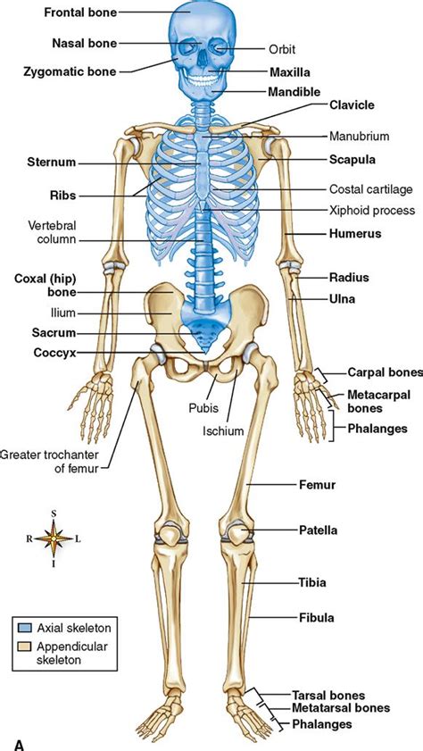
Table of Contents
The Skeletal System: A Deep Dive into the Axial and Appendicular Divisions
The human skeletal system, a marvel of biological engineering, provides the framework for our bodies. It's far more than just a collection of bones; it's a dynamic, interconnected system crucial for movement, protection of vital organs, blood cell production, and mineral storage. This intricate system is broadly divided into two main sections: the axial skeleton and the appendicular skeleton. Understanding the unique roles and components of each division is key to appreciating the overall complexity and importance of the skeletal system.
The Axial Skeleton: The Body's Central Support Structure
The axial skeleton forms the central axis of the body. Think of it as the core structure, providing support and protection for vital organs. It includes the bones of the head, neck, and trunk. This division is crucial for maintaining posture, balance, and overall body stability. Let's delve into its key components:
1. The Skull: Protecting the Brain and Sensory Organs
The skull, a complex structure of fused bones, is arguably the most important part of the axial skeleton. It's divided into two main sections:
-
Cranium: This bony enclosure protects the brain, the control center of the nervous system. It consists of several fused bones, including the frontal bone (forehead), parietal bones (top and sides of the skull), temporal bones (sides of the skull, housing the inner ear), occipital bone (back of the skull), sphenoid bone (base of the skull), and ethmoid bone (contributing to the nasal cavity and eye sockets). The intricate structure of these bones, along with their sutures (immovable joints), provides robust protection against impact.
-
Facial Bones: These bones provide the framework for the face, support the eyes, nose, and mouth, and contribute to the shape of our individual features. Key facial bones include the nasal bones (forming the bridge of the nose), zygomatic bones (cheekbones), maxillae (upper jaw), mandible (lower jaw – the only movable bone in the skull), and lacrimal bones (part of the eye socket). The intricate arrangement of these bones allows for facial expression and the intake of air and food.
2. The Vertebral Column: Flexibility and Protection for the Spinal Cord
The vertebral column, commonly known as the spine, is a flexible column of 33 vertebrae. It supports the head, neck, and trunk, while protecting the delicate spinal cord, the main pathway for nerve signals between the brain and the rest of the body. The vertebrae are grouped into five distinct regions:
-
Cervical Vertebrae (C1-C7): The seven cervical vertebrae form the neck. The first two, the atlas (C1) and axis (C2), are uniquely shaped to allow for head rotation and nodding movements.
-
Thoracic Vertebrae (T1-T12): The twelve thoracic vertebrae articulate with the ribs, forming the posterior aspect of the rib cage. This region provides support for the chest cavity and protects vital organs like the heart and lungs.
-
Lumbar Vertebrae (L1-L5): The five lumbar vertebrae are the largest and strongest, supporting the weight of the upper body. They are located in the lower back and are responsible for much of the body's flexibility in bending and twisting.
-
Sacrum: The sacrum is a triangular bone formed by the fusion of five sacral vertebrae. It connects the vertebral column to the pelvic girdle.
-
Coccyx: The coccyx, or tailbone, is the small, triangular bone formed by the fusion of three to five coccygeal vertebrae. It’s a vestigial structure, meaning it has lost much of its original function in humans.
3. The Thoracic Cage: Protecting Vital Organs
The thoracic cage, or rib cage, is formed by the twelve pairs of ribs, the sternum (breastbone), and the thoracic vertebrae. It plays a crucial role in protecting the heart, lungs, and other vital organs within the chest cavity. The ribs are connected to the sternum by costal cartilage, allowing for flexibility during breathing. The structure of the rib cage allows for expansion and contraction during respiration, facilitating efficient gas exchange.
The Appendicular Skeleton: Enabling Movement and Manipulation
The appendicular skeleton comprises the bones of the limbs and their supporting structures. This division is responsible for our ability to move around, manipulate objects, and interact with our environment. It's cleverly designed for both strength and agility. Let's examine its components:
1. The Pectoral Girdle: Connecting the Upper Limbs to the Axial Skeleton
The pectoral girdle, also known as the shoulder girdle, connects the upper limbs to the axial skeleton. It consists of the clavicles (collarbones) and scapulae (shoulder blades). These bones are relatively lightweight but provide significant flexibility, allowing for a wide range of arm movements. The unique structure of the shoulder joint, formed by the articulation of the humerus (upper arm bone) with the scapula, contributes to this remarkable mobility.
2. The Upper Limbs: Precision and Power
The upper limbs are remarkably adaptable, enabling intricate movements and powerful actions. Each upper limb comprises:
-
Humerus: The long bone of the upper arm.
-
Radius and Ulna: These two bones form the forearm, allowing for rotation of the hand and forearm.
-
Carpals, Metacarpals, and Phalanges: The carpals form the wrist; the metacarpals form the palm; and the phalanges (finger bones) allow for precise finger movements. The intricate structure of the hand allows for a wide range of dexterity and manipulative abilities, crucial for tasks requiring fine motor skills.
3. The Pelvic Girdle: Connecting the Lower Limbs to the Axial Skeleton
The pelvic girdle, or hip girdle, is a strong, stable structure formed by two hip bones (ossa coxae), which are each formed by the fusion of the ilium, ischium, and pubis. It connects the lower limbs to the axial skeleton and provides support for the abdominal organs. The pelvic girdle's robust structure is vital for bearing weight and enabling locomotion.
4. The Lower Limbs: Support and Locomotion
The lower limbs are designed for weight-bearing and locomotion. Each lower limb consists of:
-
Femur: The longest and strongest bone in the body, located in the thigh.
-
Patella: The kneecap, a sesamoid bone embedded in the quadriceps tendon, which protects the knee joint.
-
Tibia and Fibula: These two bones form the lower leg. The tibia (shinbone) is weight-bearing, while the fibula plays a role in stabilizing the ankle.
-
Tarsals, Metatarsals, and Phalanges: The tarsals form the ankle; the metatarsals form the sole of the foot; and the phalanges (toe bones) contribute to balance and propulsion during walking and running. The arch of the foot, formed by the tarsals and metatarsals, provides shock absorption and contributes to efficient locomotion.
Interconnections and Functional Importance
The axial and appendicular skeletons are not simply separate entities; they are intricately interconnected, working together to support the body's functions. The pectoral and pelvic girdles provide the crucial links between the central axis and the limbs, enabling movement and manipulation. The entire skeletal system is held together by a complex network of ligaments, tendons, and muscles, creating a dynamic, integrated system.
The skeletal system's functional importance extends far beyond support and movement:
-
Protection: The axial skeleton protects vital organs, such as the brain, spinal cord, heart, and lungs.
-
Hematopoiesis: Red and white blood cells are produced in the bone marrow, a crucial function for maintaining blood health.
-
Mineral Storage: Bones serve as a reservoir for essential minerals, such as calcium and phosphorus, which are released into the bloodstream as needed.
-
Posture and Balance: The skeletal structure plays a critical role in maintaining proper posture and balance.
-
Leverage for Movement: Bones act as levers, allowing muscles to generate force and produce movement.
Conclusion: A Remarkable System
The human skeletal system, divided into the axial and appendicular skeletons, is a testament to the intricate design of the human body. Each bone, each joint, and each connection plays a vital role in supporting life's functions. Understanding the structure and function of these two divisions offers a profound appreciation for the complexity and elegance of the human form. Further research into the specifics of bone structure, joint mechanics, and the interplay between the skeletal system and other body systems will continue to deepen our knowledge and appreciation of this remarkable biological marvel.
Latest Posts
Latest Posts
-
Graphs Of Sine And Cosine Functions Answer Key
Mar 25, 2025
-
Newspapers During The Revolutionary War Period Tended To
Mar 25, 2025
-
Are Ketones Or Aldehydes More Reactive
Mar 25, 2025
-
A Unicellular Protist Is Part Of Which Domain
Mar 25, 2025
-
A Species With 12 Protons And 10 Electrons Is
Mar 25, 2025
Related Post
Thank you for visiting our website which covers about 2 Divisions Of The Skeletal System . We hope the information provided has been useful to you. Feel free to contact us if you have any questions or need further assistance. See you next time and don't miss to bookmark.
