Basic Functional Unit Of The Kidney
Muz Play
Mar 29, 2025 · 7 min read
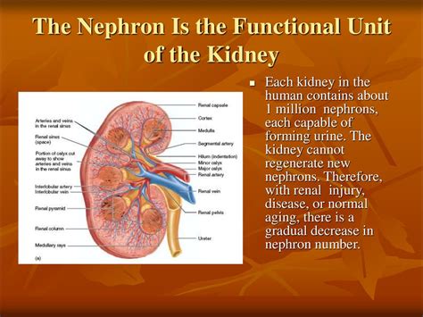
Table of Contents
The Nephron: The Basic Functional Unit of the Kidney
The human kidney, a remarkable organ, plays a vital role in maintaining homeostasis – the stable internal environment essential for survival. This intricate process is carried out by millions of microscopic units called nephrons, the basic functional units of the kidney. Understanding the nephron's structure and function is key to grasping the complexities of renal physiology and the myriad of diseases that can affect the kidneys. This article will delve deep into the structure and function of the nephron, exploring its intricate mechanisms and highlighting its importance in maintaining overall health.
The Structure of a Nephron: A Microscopic Marvel
Each nephron is a complex tubular structure, approximately 50 mm long, responsible for filtering blood and producing urine. It's comprised of two main parts: the renal corpuscle and the renal tubule.
The Renal Corpuscle: The Filtration Site
The renal corpuscle, situated in the cortex of the kidney, is the initial filtering unit. It consists of two key components:
-
Glomerulus: A network of capillaries, approximately 50-200 capillaries, where blood filtration occurs. The glomerulus is surrounded by Bowman's capsule. The specialized endothelium of the glomerular capillaries is fenestrated, meaning it's perforated with numerous pores, allowing for the passage of water and small solutes while largely preventing the passage of blood cells and large proteins. The glomerular filtration rate (GFR), the volume of fluid filtered from the glomerulus into Bowman's capsule per unit of time, is meticulously regulated to maintain homeostasis.
-
Bowman's Capsule (Glomerular Capsule): A double-walled epithelial cup that encloses the glomerulus. The inner layer, the visceral layer, consists of specialized cells called podocytes. Podocytes have long, branching processes called pedicels that interdigitate, forming filtration slits between them. These slits act as a further barrier, preventing the passage of larger proteins. The outer layer, the parietal layer, forms the outer wall of Bowman's capsule. The space between the visceral and parietal layers is called the Bowman's space, where the filtered fluid (glomerular filtrate) collects.
The Renal Tubule: Refining the Filtrate
From Bowman's capsule, the glomerular filtrate flows into the renal tubule, a long, convoluted tube responsible for modifying the filtrate's composition. The renal tubule is divided into several distinct segments:
-
Proximal Convoluted Tubule (PCT): The PCT is the longest segment of the renal tubule and is characterized by its brush border, composed of numerous microvilli, greatly increasing its surface area for reabsorption. The PCT is the site of the majority of reabsorption of essential nutrients, including glucose, amino acids, and electrolytes, as well as water. It also plays a significant role in secreting substances like hydrogen ions (H+) and ammonia (NH3). This active transport process consumes a significant amount of ATP.
-
Loop of Henle: This U-shaped segment descends into the medulla of the kidney and is crucial in establishing the medullary osmotic gradient, necessary for the concentration of urine. The descending limb is highly permeable to water but less permeable to solutes, while the ascending limb is impermeable to water but actively transports sodium, potassium, and chloride ions out of the tubule. This countercurrent multiplier system allows for the efficient reabsorption of water and the creation of a hypertonic medullary interstitium. The length of the Loop of Henle varies depending on the nephron's location and function. Juxtamedullary nephrons have significantly longer loops of Henle than cortical nephrons, contributing to their role in urine concentration.
-
Distal Convoluted Tubule (DCT): The DCT is the final segment of the renal tubule and is regulated by hormones such as aldosterone and parathyroid hormone. Aldosterone, released by the adrenal glands, stimulates sodium reabsorption and potassium secretion, influencing blood pressure and electrolyte balance. Parathyroid hormone promotes calcium reabsorption and phosphate excretion. The DCT is also involved in fine-tuning the reabsorption and secretion of ions to maintain the body's fluid and electrolyte balance.
-
Collecting Duct: While not technically part of the nephron, the collecting duct is vital in the final processing of urine. Several nephrons drain into a single collecting duct. The collecting ducts are also under hormonal control, primarily by antidiuretic hormone (ADH). ADH increases water reabsorption in the collecting ducts, leading to the production of concentrated urine.
The Function of a Nephron: Filtration, Reabsorption, and Secretion
The nephron's function is summarized by three critical processes: filtration, reabsorption, and secretion.
Glomerular Filtration: The Initial Step
Glomerular filtration is a passive process driven by hydrostatic pressure differences across the glomerular capillaries. The high blood pressure in the glomerulus forces water and small solutes across the filtration barrier into Bowman's space, forming the glomerular filtrate. Large molecules, such as proteins and blood cells, are largely excluded. The filtration process is highly efficient, with approximately 180 liters of filtrate produced per day.
Tubular Reabsorption: Reclaiming Essentials
Tubular reabsorption is the selective retrieval of essential substances from the glomerular filtrate back into the bloodstream. This process is both active (requiring energy) and passive (driven by concentration gradients). Glucose, amino acids, and many electrolytes are almost completely reabsorbed in the PCT. Water reabsorption occurs throughout the renal tubule, primarily driven by osmosis in response to solute reabsorption. The Loop of Henle plays a vital role in water reabsorption via the countercurrent mechanism. The DCT and collecting duct contribute to fine-tuning electrolyte and water balance.
Tubular Secretion: Eliminating Waste
Tubular secretion is the active transport of certain substances from the bloodstream into the renal tubule. This process helps eliminate waste products, such as toxins, drugs, and excess ions, that were not filtered into Bowman's space or that were reabsorbed. Hydrogen ions (H+) and potassium ions (K+) are actively secreted in the DCT, contributing to acid-base balance and electrolyte homeostasis.
Types of Nephrons: Cortical and Juxtamedullary
Nephrons are broadly classified into two types based on their location and the length of their Loop of Henle:
-
Cortical Nephrons: These nephrons are the most abundant type, representing about 85% of the total nephron population. Their renal corpuscles are located in the outer cortex, and their Loops of Henle are relatively short, extending only slightly into the medulla. Cortical nephrons play a major role in the reabsorption of nutrients and electrolytes.
-
Juxtamedullary Nephrons: These nephrons have their renal corpuscles located close to the corticomedullary junction, and their Loops of Henle extend deep into the medulla. The long loops of Henle are crucial in establishing the medullary osmotic gradient, which is essential for concentrating urine. These nephrons contribute significantly to the kidney's ability to conserve water.
Juxtaglomerular Apparatus (JGA): Regulation of Blood Pressure and GFR
The juxtaglomerular apparatus (JGA) is a specialized structure located where the distal convoluted tubule contacts the afferent and efferent arterioles of the glomerulus. The JGA plays a vital role in regulating glomerular filtration rate (GFR) and blood pressure. It comprises three cell types:
-
Juxtaglomerular Cells: These specialized smooth muscle cells in the afferent arteriole are responsible for secreting renin, an enzyme that plays a key role in the renin-angiotensin-aldosterone system (RAAS). Renin initiates a cascade of events leading to increased blood pressure and sodium reabsorption.
-
Macula Densa Cells: These are specialized epithelial cells in the distal convoluted tubule that monitor the concentration of sodium chloride (NaCl) in the filtrate. They respond to changes in GFR, influencing renin release from the juxtaglomerular cells.
-
Extraglomerular Mesangial Cells: These cells act as intercellular messengers between the macula densa and juxtaglomerular cells.
Clinical Significance: Renal Diseases and Nephron Damage
Damage to nephrons, resulting from various diseases, leads to impaired kidney function. Conditions such as glomerulonephritis (inflammation of the glomeruli), diabetic nephropathy (kidney damage caused by diabetes), and polycystic kidney disease (formation of cysts in the kidneys) all affect nephron structure and function, leading to decreased GFR and ultimately, kidney failure. Understanding the structure and function of the nephron is crucial for diagnosing and managing renal diseases.
Conclusion: The Nephron's Indispensable Role
The nephron, the basic functional unit of the kidney, is a marvel of biological engineering. Its intricate structure and precise functions enable the kidney to maintain homeostasis by carefully regulating the composition of body fluids. The nephron's involvement in filtration, reabsorption, and secretion is vital for removing waste products, maintaining electrolyte balance, regulating blood pressure, and controlling blood volume. Comprehensive understanding of nephron structure and function is critical not only for appreciating renal physiology but also for understanding the pathogenesis and treatment of numerous renal diseases. Further research continues to unravel the intricacies of nephron function and its contribution to overall health and well-being. Protecting these vital microscopic units through healthy lifestyle choices is paramount for long-term kidney health.
Latest Posts
Latest Posts
-
Provide The Correct Systematic Name For The Compound Shown Here
Apr 01, 2025
-
Are All Ionic Compounds Strong Electrolytes
Apr 01, 2025
-
How Do You Use A Balance
Apr 01, 2025
-
Nucleotide Excision Repair Vs Mismatch Repair
Apr 01, 2025
-
What Is The Difference Between Religious And Ethnic Groups
Apr 01, 2025
Related Post
Thank you for visiting our website which covers about Basic Functional Unit Of The Kidney . We hope the information provided has been useful to you. Feel free to contact us if you have any questions or need further assistance. See you next time and don't miss to bookmark.
