Cell Bodies Of Sensory Neurons Are Located In
Muz Play
Mar 17, 2025 · 6 min read
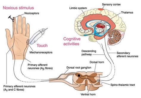
Table of Contents
Cell Bodies of Sensory Neurons: Location and Significance
The intricate human nervous system relies on a complex interplay of neurons to transmit information throughout the body. Sensory neurons, a crucial component of this system, are responsible for detecting stimuli from the internal and external environment and relaying this information to the central nervous system (CNS). Understanding the location of the cell bodies of these sensory neurons is fundamental to comprehending how sensory information is processed and relayed. This article will delve deep into the precise locations of sensory neuron cell bodies, exploring the different types of sensory neurons and their associated pathways.
The Diverse Locations of Sensory Neuron Cell Bodies
Unlike motor neurons, whose cell bodies are predominantly located within the CNS, sensory neuron cell bodies are situated outside the CNS in various ganglia. These ganglia act as relay stations, organizing and processing sensory information before it reaches the brain and spinal cord. The location of these ganglia varies significantly depending on the type of sensory information being relayed.
1. Dorsal Root Ganglia (DRG): The Home of Somatic Sensory Neurons
The most prominent location for sensory neuron cell bodies is the dorsal root ganglia (DRG). These ganglia are located along the spinal cord, with a pair associated with each spinal nerve. The DRG house the cell bodies of somatic sensory neurons, which receive sensory input from the skin, muscles, joints, and other somatic structures. These neurons are responsible for detecting a wide range of sensations, including:
- Touch: Light touch, pressure, vibration.
- Temperature: Heat and cold.
- Pain: Nociception (detection of harmful stimuli).
- Proprioception: Awareness of body position and movement.
The arrangement within the DRG is remarkably organized. Each DRG contains thousands of neuronal cell bodies, each corresponding to a specific sensory receptor in the periphery. The axons of these neurons extend peripherally to their respective receptors, and centrally into the spinal cord, forming the dorsal roots. This arrangement ensures efficient transmission of somatic sensory information to the spinal cord for further processing and relay to the brain.
Further Exploration of Somatic Sensory Neuron Pathways: Understanding the pathways followed by different types of somatic sensory information is crucial. For instance, touch information often follows different pathways than pain and temperature information, leading to different processing centers in the brain. The specific receptors involved also influence the pathway. Mechanoreceptors involved in touch sensation have different fiber types and conduction velocities compared to nociceptors sensing pain. This difference influences the speed of response and the perception of the stimulus.
2. Cranial Nerve Ganglia: Sensory Input to the Brain
The cranial nerves, twelve pairs of nerves emerging directly from the brainstem, also contain sensory neuron cell bodies located in associated ganglia. These ganglia are named according to the respective cranial nerve, such as:
- Trigeminal ganglion: This ganglion houses the cell bodies of sensory neurons associated with the trigeminal nerve (CN V), which innervates the face, head, and oral cavity. These neurons are responsible for detecting touch, temperature, pain, and proprioception in the face.
- Vestibulocochlear ganglion: Located within the inner ear, this ganglion contains the cell bodies of sensory neurons associated with the vestibulocochlear nerve (CN VIII), responsible for hearing and balance. These neurons detect sound waves and head movements.
- Geniculate ganglion: Associated with the facial nerve (CN VII), this ganglion contains cell bodies of sensory neurons involved in taste sensation and somatosensation in parts of the face.
- Glossopharyngeal ganglion: Associated with the glossopharyngeal nerve (CN IX), this ganglion holds cell bodies of neurons responsible for taste and somatosensation in the posterior tongue and pharynx.
- Superior and Inferior Ganglia of the Vagus Nerve: These ganglia contain cell bodies of sensory neurons associated with the vagus nerve (CN X), relaying visceral sensory information from the thorax and abdomen.
The sensory information processed by these ganglia is crucial for various essential functions, including facial expression, hearing, balance, and autonomic regulation.
Detailed Pathway Analysis: The pathways from these cranial nerve ganglia to specific regions of the brainstem and thalamus vary depending on the type of sensory information. This anatomical complexity underscores the specialized nature of sensory processing in the brain. Each pathway is precisely mapped, allowing for accurate interpretation of the sensory stimuli.
3. Autonomic Ganglia: Sensory Input from Viscera
The autonomic nervous system regulates involuntary functions like heart rate, digestion, and respiration. Sensory neurons within this system, known as visceral sensory neurons, monitor these internal organs and relay information to the CNS. Their cell bodies are located in various autonomic ganglia, including:
- Prevertebral ganglia: Located anterior to the vertebral column, these ganglia receive sensory input from the abdominal viscera.
- Paravertebral ganglia (sympathetic chain): Situated along the vertebral column, these ganglia receive sensory input related to sympathetic innervation.
- Terminal ganglia: These ganglia are located near or within the target organs.
Visceral sensory information differs from somatic sensation in several key aspects. The sensitivity is often less precise, and the perception is often less localized. Pain sensations from internal organs can be referred to other parts of the body, a phenomenon known as referred pain. This is due to the convergence of visceral and somatic sensory afferents on the same neurons in the spinal cord.
4. Other Sensory Neuron Locations
While the DRG, cranial nerve ganglia, and autonomic ganglia are the primary locations, other locations house a smaller number of sensory neuron cell bodies. These include specialized ganglia associated with certain sensory organs, such as the olfactory bulb for smell and the retina for vision. These specialized ganglia contribute to specific sensory pathways with unique characteristics.
The Functional Significance of Sensory Neuron Cell Body Location
The location of sensory neuron cell bodies isn't arbitrary. Their peripheral position has several crucial implications:
-
Efficient Signal Transmission: Placing the cell bodies outside the CNS allows for rapid signal transmission across shorter distances. Sensory information is processed closer to the sensory receptors, allowing for faster reflexes and immediate responses to stimuli.
-
Protection: The ganglia act as protective sheaths, shielding the cell bodies from damage. The surrounding connective tissue and glial cells provide a supportive environment, preventing direct exposure to potentially harmful stimuli.
-
Organization and Processing: Ganglia act as relay stations, allowing for the organization and initial processing of sensory information before it reaches the CNS. This reduces the processing load on the central nervous system and allows for more efficient information handling.
-
Modulation of Sensory Input: Sensory input can be modulated at the level of the ganglia through various mechanisms, including synaptic transmission and neurotransmitter interactions. This provides a level of control over sensory perception and responsiveness.
Conclusion: A Complex Network of Sensory Perception
The location of sensory neuron cell bodies is a critical aspect of the intricate sensory processing system. The precise placement within DRG, cranial nerve ganglia, autonomic ganglia, and other specialized locations reflects the complexity of sensory input and the need for efficient and organized relay of information to the CNS. Understanding the location and function of these ganglia is vital for comprehending the mechanisms of sensation, perception, and reflexive responses. Further research continues to elucidate the intricacies of sensory neuron organization and the molecular mechanisms governing their function, offering insights into treating sensory disorders and improving our understanding of the nervous system. The field is dynamic, with ongoing advancements in our understanding of the detailed pathways and processing at each stage. The study of sensory neuron location and function provides a foundation for research into chronic pain conditions, neurodegenerative diseases, and the development of new therapeutic strategies.
Latest Posts
Latest Posts
-
What Is The Base Of A Triangle
Mar 17, 2025
-
Electrons Are Located In Energy Levels Called Electron
Mar 17, 2025
-
Can Mitochondria Survive Outside The Cell
Mar 17, 2025
-
Name A Structural Difference Between Triglycerides And Phospholipids
Mar 17, 2025
-
Ending Materials In A Chemical Reaction
Mar 17, 2025
Related Post
Thank you for visiting our website which covers about Cell Bodies Of Sensory Neurons Are Located In . We hope the information provided has been useful to you. Feel free to contact us if you have any questions or need further assistance. See you next time and don't miss to bookmark.
