Clusters Of Neuron Cell Bodies In The Pns Are Called
Muz Play
Mar 31, 2025 · 5 min read
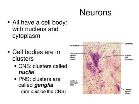
Table of Contents
Clusters of Neuron Cell Bodies in the PNS are Called Ganglia: A Deep Dive into Peripheral Nervous System Organization
Clusters of neuron cell bodies in the peripheral nervous system (PNS) are called ganglia. Understanding ganglia is crucial to comprehending the intricate workings of the PNS, its role in communication between the central nervous system (CNS) and the body's periphery, and the various pathologies that can affect this vital system. This article will delve into the detailed anatomy, function, types, and clinical significance of ganglia.
What are Ganglia?
Ganglia are collections of neuronal cell bodies, also known as somata, located outside the CNS. They serve as relay stations, integrating sensory information from the periphery and transmitting signals to and from the CNS. Unlike the gray matter of the brain and spinal cord, which also contains neuronal cell bodies, ganglia are distinct structures within the PNS, often enveloped by connective tissue for protection and structural integrity. This connective tissue sheath provides structural support and separates the ganglia from surrounding tissues.
Key Differences Between Ganglia and Nuclei:
It's important to distinguish ganglia from nuclei. While both are collections of neuronal cell bodies, their location defines the key difference:
- Ganglia: Located in the PNS.
- Nuclei: Located within the CNS (brain and spinal cord).
Types of Ganglia in the Peripheral Nervous System
The PNS boasts diverse ganglia, each playing a specific role. These can be broadly categorized based on their function and location:
1. Sensory Ganglia (Posterior Root Ganglia or Dorsal Root Ganglia):
These ganglia are associated with sensory neurons, relaying sensory information from the periphery to the CNS. They are strategically located along the dorsal roots of spinal nerves, hence their alternative names. The cell bodies of sensory neurons, which have long processes extending to the periphery and into the spinal cord, reside within these ganglia. These sensory neurons are pseudounipolar, meaning they have a single axon that splits into two branches: one extending to the periphery to receive sensory stimuli (e.g., touch, pain, temperature) and another extending to the spinal cord to transmit the signal.
Sensory Neuron Function within Dorsal Root Ganglia:
The sensory neurons within the dorsal root ganglia are highly specialized, responsible for:
- Somatosensation: Detecting sensations from the skin, muscles, and joints.
- Viscerosensation: Detecting sensations from internal organs.
2. Autonomic Ganglia:
These ganglia are part of the autonomic nervous system (ANS), which controls involuntary functions such as heart rate, digestion, and respiration. They are crucial in the relay of signals from the CNS to target organs (effectors). The ANS further divides into the sympathetic and parasympathetic nervous systems, each having its own distinct ganglionic organization:
a) Sympathetic Ganglia:
These ganglia form the sympathetic trunk, a chain of ganglia running alongside the vertebral column. Preganglionic sympathetic neurons, originating in the spinal cord, synapse with postganglionic neurons within these ganglia. The postganglionic neurons then innervate various organs, preparing the body for "fight-or-flight" responses.
Sympathetic Ganglia Features:
- Paravertebral location: Positioned alongside the vertebral column.
- Short preganglionic fibers, long postganglionic fibers: This arrangement allows for widespread effects.
- Release of norepinephrine: The primary neurotransmitter at the postganglionic synapse.
b) Parasympathetic Ganglia:
Unlike the sympathetic ganglia's chain-like structure, parasympathetic ganglia are located near or within the target organs they innervate. Preganglionic parasympathetic fibers, originating from the brainstem and sacral spinal cord, extend to these ganglia, forming synapses with postganglionic neurons. These postganglionic neurons then modulate organ function, promoting "rest-and-digest" responses.
Parasympathetic Ganglia Features:
- Terminal ganglia: Located near or within the target organs.
- Long preganglionic fibers, short postganglionic fibers: Allows for more localized effects.
- Release of acetylcholine: The primary neurotransmitter at the postganglionic synapse.
3. Enteric Ganglia:
These ganglia form the enteric nervous system (ENS), a complex network of neurons embedded within the walls of the gastrointestinal tract. The ENS is often referred to as the "second brain" because of its extensive capacity for independent function. It controls various gastrointestinal processes, including motility, secretion, and absorption, with minimal influence from the CNS.
Enteric Ganglia Function:
- Gut motility: Coordinates muscle contractions for food propulsion.
- Secretion: Regulates the release of digestive enzymes and hormones.
- Absorption: Influences the uptake of nutrients.
Clinical Significance of Ganglia
Several clinical conditions can directly or indirectly affect ganglia, leading to diverse symptoms and functional impairments:
1. Ganglion Cysts:
These are benign, fluid-filled sacs that commonly develop near joints, often in the hands and wrists. Although not directly involving the ganglia described above, the term "ganglion" is used due to their appearance as a localized swelling. The exact cause of ganglion cysts is unknown, but they may be associated with repetitive movements or joint injury.
2. Shingles (Herpes Zoster):
This viral infection affects sensory ganglia, specifically the dorsal root ganglia, resulting in a painful rash along the affected dermatome (area of skin innervated by a single sensory nerve). The varicella-zoster virus, the same virus that causes chickenpox, reactivates in the ganglia, causing inflammation and nerve damage.
3. Autonomic Neuropathies:
These conditions involve damage to the autonomic nervous system, potentially affecting autonomic ganglia. Diabetes mellitus is a common cause, leading to impaired function in various organs. Symptoms can include orthostatic hypotension, gastrointestinal disturbances, and bladder dysfunction.
4. Neuroblastoma:
This is a rare cancer arising from immature nerve cells, often originating in the adrenal medulla or sympathetic ganglia. It's more common in infants and young children.
5. Guillain-Barré Syndrome:
This autoimmune disorder attacks the myelin sheath surrounding peripheral nerves, potentially affecting sensory and autonomic ganglia. It can lead to muscle weakness, paralysis, and autonomic dysfunction.
Research and Future Directions
Ongoing research continues to explore the intricate complexities of ganglia, focusing on:
- Development and differentiation of ganglionic neurons: Understanding the molecular mechanisms that govern their formation during embryonic development.
- Role of ganglia in pain processing: Investigating the contribution of ganglia to chronic pain conditions.
- Therapeutic targeting of ganglia: Developing novel treatments for disorders affecting ganglionic function.
Conclusion
Ganglia, clusters of neuronal cell bodies in the PNS, play a pivotal role in relaying sensory information, controlling involuntary functions, and coordinating gastrointestinal processes. Their diverse types and locations reflect the complexity of the peripheral nervous system. A thorough understanding of ganglionic anatomy, function, and associated pathologies is crucial for diagnosing and treating a broad spectrum of neurological and other medical conditions. Further research will undoubtedly reveal even more about the intricate role of ganglia in maintaining the overall health and well-being of the body. Understanding these crucial components of the nervous system offers invaluable insights into both normal physiology and the pathophysiology of numerous diseases.
Latest Posts
Latest Posts
-
How To Make Normal Probability Plot
Apr 02, 2025
-
Chemical Kinetics Of The Iodine Clock Reaction Lab Report
Apr 02, 2025
-
Who Is Credited With Discovering Cells
Apr 02, 2025
-
Calculate The Ph At The Equivalence Point For The Titration
Apr 02, 2025
-
Gramatica A The Verb Tener Answers
Apr 02, 2025
Related Post
Thank you for visiting our website which covers about Clusters Of Neuron Cell Bodies In The Pns Are Called . We hope the information provided has been useful to you. Feel free to contact us if you have any questions or need further assistance. See you next time and don't miss to bookmark.
