Coleus Stem Tip Under Microscope Labeled
Muz Play
Mar 25, 2025 · 6 min read
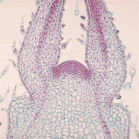
Table of Contents
Coleus Stem Tip Under the Microscope: A Detailed Exploration
Coleus, with its vibrant and diverse leaf colors, is a popular choice for gardeners and a fascinating subject for microscopic study. Observing a Coleus stem tip under a microscope reveals a complex world of cellular structures and processes essential for plant growth and development. This article delves into the intricate details of a Coleus stem tip's microscopic anatomy, exploring its key features and their significance. We'll cover everything from identifying specific cell types to understanding the implications of observing these structures.
Preparing the Coleus Stem Tip for Microscopic Observation
Before we delve into the intricacies of the microscopic view, let's address the crucial first step: sample preparation. The success of your microscopic observation hinges on proper preparation techniques. Here's a step-by-step guide:
1. Selecting the Sample:
Choose a young, actively growing Coleus stem tip. The tender, rapidly dividing cells of a young stem provide clearer visualization of cellular structures. Avoid stems that are woody or overly mature.
2. Sectioning the Sample:
Obtain thin, translucent sections of the stem tip. This can be achieved using a sharp razor blade or a microtome (for thinner, more consistent sections). Extremely thin sections (around 5-10 micrometers) are optimal for light microscopy. The goal is to create sections thin enough to allow light to pass through, enabling clear visualization of cellular details.
3. Staining (Optional):
Staining enhances the visibility of cellular components. Common stains for plant tissues include:
- Iodine: A simple stain that highlights cell walls.
- Methylene blue: Stains cell nuclei and other structures.
- Eosin: A counterstain often used in combination with methylene blue, highlighting different cellular components with contrasting colors.
The choice of stain depends on the specific structures you wish to highlight. Remember to follow the stain's instructions carefully.
4. Mounting the Sample:
Once stained (if applicable), carefully mount the section onto a microscope slide using a mounting medium, such as glycerin or water. A coverslip should then be gently placed over the sample to protect it and flatten it for better viewing. Avoid trapping air bubbles, which can obstruct the view.
Microscopic Anatomy of the Coleus Stem Tip: A Detailed Look
Now, let's explore the fascinating cellular structures visible under the microscope.
1. Epidermis:
The outermost layer of cells, the epidermis, forms a protective barrier against the environment. Under the microscope, you'll observe a single layer of tightly packed, relatively flat cells. These cells often have a slightly irregular shape and may possess a cuticle, a waxy layer that reduces water loss. The epidermis plays a vital role in preventing water loss, regulating gas exchange, and protecting the underlying tissues.
2. Cortex:
Beneath the epidermis lies the cortex, consisting primarily of parenchyma cells. These are relatively large, thin-walled cells with a variety of functions, including storage of food reserves and support. In a Coleus stem, you might observe different types of parenchyma cells, potentially including chlorenchyma (containing chloroplasts for photosynthesis) and storage parenchyma. The arrangement of these cells can provide structural support for the stem.
3. Vascular Bundles:
A defining feature of the Coleus stem is its vascular bundles, responsible for the transport of water, minerals, and sugars. These bundles appear as distinct, elongated structures under the microscope. Each vascular bundle contains:
- Xylem: Conducts water and minerals from the roots to the leaves. Xylem cells are typically elongated, thick-walled, and often appear empty (hollow) under the microscope, reflecting their function in water transport. You may observe different types of xylem cells, including tracheids and vessel elements.
- Phloem: Conducts sugars (produced during photosynthesis) from the leaves to other parts of the plant. Phloem cells are also elongated but often appear smaller and less clearly defined than xylem cells under the microscope. Sieve tubes and companion cells are key components of the phloem. These cells work together to facilitate efficient sugar transport.
The arrangement of the xylem and phloem can vary depending on the age and type of plant; in Coleus, you'll likely see a distinct arrangement, often described as a collateral vascular bundle (xylem and phloem adjacent to each other).
4. Pith:
The central core of the stem is the pith, primarily composed of parenchyma cells. In Coleus, the pith may be relatively large and contain loosely packed parenchyma cells. The pith provides support and stores nutrients.
5. Intercellular Spaces:
Between the cells in the cortex and pith, you'll likely observe intercellular spaces—gaps between the cells. These spaces facilitate gas exchange and allow for flexibility and growth. The size and distribution of these spaces can vary across different tissue types.
Identifying Specific Cell Types and Structures
Detailed identification relies heavily on staining and magnification. However, here are some key features to observe under the microscope to aid in identification:
- Cell Walls: Look for the presence and thickness of cell walls. The primary cell wall is usually thin, while secondary cell walls (present in some xylem cells) are much thicker and more rigid.
- Cell Nuclei: Staining with dyes like methylene blue can help to highlight the nuclei, revealing the size, shape, and location of the nuclei within the cells.
- Chloroplasts: In chlorenchyma cells, chloroplasts will appear as small, green organelles within the cytoplasm. Their presence indicates the cells' role in photosynthesis.
- Sieve Plates: In phloem sieve tube elements, look for sieve plates, specialized areas where the cell walls have perforations that facilitate the flow of sugars.
Interpreting Your Observations: Growth and Development
Microscopic observations of the Coleus stem tip provide valuable insights into plant growth and development. Here’s how:
- Apical Meristem: The very tip of the stem houses the apical meristem, a region of actively dividing cells responsible for primary growth (increase in stem length). The high density of these cells and their visible mitotic activity (cell division) are clear indicators of this region's importance in growth.
- Cell Differentiation: As cells move away from the apical meristem, they differentiate into specialized cell types (epidermis, parenchyma, xylem, phloem). Observing this transition provides a visual representation of plant cell differentiation.
- Vascular Development: The arrangement and development of vascular bundles indicate the plant's efficiency in transporting water and nutrients. Changes in the size and complexity of these bundles reflect growth and development.
- Effect of Environmental Factors: Microscopic studies can reveal the effects of environmental stresses (e.g., drought, nutrient deficiency) on plant cell structure and function.
Beyond the Basic Observation: Advanced Techniques
While light microscopy provides a fundamental understanding of Coleus stem tip anatomy, more advanced techniques offer even greater detail:
- Electron Microscopy (TEM and SEM): These techniques offer significantly higher resolution, allowing for the visualization of ultrastructural details (e.g., membranes, organelles) not visible with light microscopy.
- Immunohistochemistry: This technique uses antibodies to label specific proteins within the cells, allowing researchers to pinpoint the location and abundance of particular molecules involved in plant growth and development.
- Confocal Microscopy: Confocal microscopy provides three-dimensional images of plant tissues, allowing for a more comprehensive understanding of cellular arrangements.
Conclusion: The Value of Microscopic Study
The microscopic study of a Coleus stem tip is a rewarding endeavor, revealing the complex and intricate cellular organization underlying plant growth and development. From the simple identification of cell types to the investigation of advanced physiological processes, this observation provides a powerful tool for learning about plant biology. By combining careful sample preparation, appropriate staining, and keen observation, researchers and students can unlock the secrets hidden within this seemingly simple plant stem. The knowledge gained from such microscopic studies contributes significantly to our understanding of plant biology, ultimately helping us address important challenges in agriculture, horticulture, and environmental science.
Latest Posts
Latest Posts
-
What Is The Radius Of Hydrogen
Mar 28, 2025
-
Is Trigonal Planar Polar Or Nonpolar
Mar 28, 2025
-
What Is The Unique Property Of Water
Mar 28, 2025
-
What Is A Particle With A Negative Charge
Mar 28, 2025
-
Why Does Water Have High Heat Of Vaporization
Mar 28, 2025
Related Post
Thank you for visiting our website which covers about Coleus Stem Tip Under Microscope Labeled . We hope the information provided has been useful to you. Feel free to contact us if you have any questions or need further assistance. See you next time and don't miss to bookmark.
