Color A Typical Prokaryote Cell Answer Key
Muz Play
Mar 30, 2025 · 5 min read
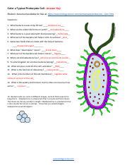
Table of Contents
Coloring a Typical Prokaryote Cell: A Detailed Guide with Answer Key
Understanding prokaryotic cell structure is fundamental to grasping the basics of biology. This comprehensive guide will walk you through the process of coloring a typical prokaryote cell, providing a detailed explanation of each structure and its function. We'll also provide an answer key to help you verify your work and solidify your understanding. This guide incorporates various learning techniques to ensure effective knowledge retention.
What is a Prokaryotic Cell?
Before we dive into the coloring exercise, let's refresh our understanding of prokaryotic cells. Prokaryotes are single-celled organisms lacking a membrane-bound nucleus and other membrane-bound organelles. This contrasts sharply with eukaryotic cells, which possess a nucleus and various other organelles. Prokaryotes are incredibly diverse, encompassing bacteria and archaea, and play crucial roles in various ecosystems.
Key Structures of a Prokaryotic Cell:
To accurately color your prokaryotic cell, you need to understand the key structures and their functions:
1. Cell Wall: This rigid outer layer provides structural support and protection. In bacteria, the cell wall typically contains peptidoglycan, a unique polymer. Color suggestion: Dark Brown
2. Plasma Membrane (Cell Membrane): Located beneath the cell wall, this selectively permeable membrane regulates the passage of substances into and out of the cell. Color suggestion: Light Blue
3. Cytoplasm: The jelly-like substance filling the cell, containing ribosomes and the genetic material. Color suggestion: Light Green
4. Nucleoid: This is the region where the prokaryotic DNA is located. Unlike a eukaryotic nucleus, it is not membrane-bound. Color suggestion: Purple
5. Ribosomes: These are the sites of protein synthesis. They are smaller than eukaryotic ribosomes. Color suggestion: Dark Green
6. Plasmids (Optional): These are small, circular DNA molecules separate from the chromosomal DNA. They often carry genes that provide advantages, such as antibiotic resistance. Color suggestion: Pink
7. Flagella (Optional): These are long, whip-like appendages used for locomotion. Not all prokaryotes possess flagella. Color suggestion: Red
8. Pili (Optional): These are short, hair-like appendages involved in attachment to surfaces and bacterial conjugation (transfer of genetic material). Color suggestion: Orange
9. Capsule (Optional): A sticky, outer layer found in some bacteria; it provides protection and aids in attachment to surfaces. Color suggestion: Light Brown
Coloring Exercise: A Step-by-Step Guide
-
Obtain Your Materials: You'll need a drawing of a typical prokaryotic cell (either pre-drawn or one you create), colored pencils or crayons, and a sharpener.
-
Labeling is Key: Before you start coloring, carefully label the major structures on your drawing. This helps reinforce your understanding of the cell's components.
-
Strategic Coloring: Following the color suggestions above (or choosing your own!), begin coloring each structure. Pay attention to the relative sizes and positions of these structures within the cell.
-
Adding Detail (Optional): Once you've colored the basic structures, you can add further detail to enhance your diagram. This could include shading to give a sense of depth or drawing the structures at different stages of activity (for example, showing flagella in motion).
-
Accuracy Matters: Ensure your coloring accurately reflects the location and arrangement of cell components. A well-colored diagram should clearly differentiate the various parts of the prokaryotic cell.
Answer Key & Structure Verification:
After completing your colored diagram, compare your work to the descriptions below. This will help you check for accuracy and reinforce your knowledge.
-
Cell Wall (Dark Brown): Should be the outermost layer, surrounding the entire cell, providing a clear boundary.
-
Plasma Membrane (Light Blue): Should be located immediately beneath the cell wall, depicted as a thin layer.
-
Cytoplasm (Light Green): Should fill the cell, occupying the space between the cell membrane and the nucleoid.
-
Nucleoid (Purple): Should be located within the cytoplasm, often depicted as a less distinct, irregularly shaped region.
-
Ribosomes (Dark Green): Should be scattered throughout the cytoplasm, represented as small dots or granules.
-
Plasmids (Pink, Optional): If included, should be depicted as small, circular structures within the cytoplasm.
-
Flagella (Red, Optional): If included, should be long, whip-like structures extending from the cell's surface.
-
Pili (Orange, Optional): If included, should be shorter and thinner than flagella, often multiple appendages.
-
Capsule (Light Brown, Optional): If included, should be the outermost layer, encompassing the cell wall and giving a fuzzy appearance.
Beyond Coloring: Deeper Understanding
Coloring the prokaryotic cell is a starting point for understanding its complex structure and function. To enhance your knowledge, consider these additional points:
-
Differences between Bacteria and Archaea: While both are prokaryotes, they differ significantly in their cell wall composition and other molecular features.
-
Bacterial Morphology: Bacteria exhibit various shapes, including cocci (spherical), bacilli (rod-shaped), and spirilla (spiral-shaped). Understanding these morphologies is crucial for identification and classification.
-
Prokaryotic Metabolism: Prokaryotes display a remarkable diversity in their metabolic pathways, including aerobic respiration, anaerobic respiration, fermentation, and photosynthesis.
-
Prokaryotic Genetics: Studying prokaryotic genetics reveals insights into gene regulation, mutation, and horizontal gene transfer.
Advanced Activities:
-
Comparative Analysis: Create a comparative diagram of prokaryotic and eukaryotic cells, highlighting their similarities and differences.
-
Research Specific Bacteria: Investigate the structure and function of specific bacterial species, paying attention to their unique adaptations.
-
Illustrative Essays: Write short essays explaining the roles of specific prokaryotic structures in cell survival and function.
This detailed guide offers a structured approach to coloring a typical prokaryotic cell, going beyond a simple coloring exercise to foster a deeper understanding of its complexity. Remember, the key is to understand the function of each structure and its place within the cell’s overall organization. This activity provides a strong foundation for further exploration of microbiology and the fascinating world of prokaryotic cells. Through diligent study and application of these techniques, you can confidently master the intricacies of this fundamental biological topic.
Latest Posts
Latest Posts
-
How To Calculate Standard Free Energy Change
Apr 01, 2025
-
Root Mean Square Velocity Of Gas
Apr 01, 2025
-
Where Is The Respiratory Center Located In The Brain
Apr 01, 2025
-
What Is The Difference Between Cellular Respiration And Fermentation
Apr 01, 2025
-
What Are Two Functional Groups Found In Amino Acids
Apr 01, 2025
Related Post
Thank you for visiting our website which covers about Color A Typical Prokaryote Cell Answer Key . We hope the information provided has been useful to you. Feel free to contact us if you have any questions or need further assistance. See you next time and don't miss to bookmark.
