Cross Section Of A Leaf Microscope
Muz Play
Mar 20, 2025 · 7 min read
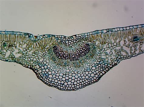
Table of Contents
A Deep Dive into the Cross Section of a Leaf Under the Microscope: Unveiling the Secrets of Photosynthesis
The humble leaf, often overlooked in the grandeur of the plant kingdom, is a powerhouse of biological processes. At its core lies a complex structure meticulously designed for photosynthesis, the vital process that converts sunlight into energy. Exploring the cross-section of a leaf under a microscope reveals a captivating world of cellular architecture, perfectly adapted for this crucial function. This detailed exploration will guide you through the intricacies of leaf anatomy, highlighting key features and their roles in photosynthesis and overall plant health.
Preparing the Leaf for Microscopic Examination: A Step-by-Step Guide
Before embarking on our microscopic journey, preparing the leaf sample is crucial. The process ensures optimal visualization of the internal structures. Here's a simple yet effective method:
1. Sample Selection:
Choose a healthy, mature leaf from a readily available plant. Avoid leaves that show signs of disease or damage. Dicots (plants with two cotyledons) are excellent choices because their leaves exhibit a clear venation pattern, making observation easier.
2. Fixation:
This step preserves the leaf's cellular structure by halting enzymatic activity. Immerse the leaf in a fixative solution (e.g., formalin or FAA - Formaldehyde-Acetic Acid-Alcohol) for a few hours, or preferably overnight.
3. Dehydration:
Remove excess water from the leaf tissue using a graded ethanol series (increasing concentrations of ethanol in water, e.g., 30%, 50%, 70%, 90%, 100%). This prepares the leaf for embedding in paraffin wax.
4. Embedding:
Infiltrate the leaf with molten paraffin wax to provide support and facilitate sectioning. Allow the wax to solidify completely.
5. Sectioning:
Using a microtome, carefully cut thin sections (ideally 8-10 µm thick) of the embedded leaf. These thin sections are essential for light microscopy, as they allow light to pass through, allowing visualization of internal structures.
6. Mounting and Staining:
Mount the sections onto glass slides using a mounting medium (e.g., DPX). Stain the sections with appropriate stains (e.g., toluidine blue or safranin) to enhance contrast and visualize different cell types and structures.
Key Structures Revealed Under the Microscope: A Microscopic Tour
With the prepared slide under the microscope, we can now explore the key anatomical features that contribute to the leaf's remarkable functionality.
1. The Epidermis: A Protective Shield
The upper epidermis, the outermost layer on the leaf's upper surface, is typically a single layer of transparent cells, forming a protective barrier against water loss and pathogen invasion. The cuticle, a waxy layer covering the epidermis, significantly reduces water loss through transpiration.
Specialized Epidermal Cells: The epidermis may also contain specialized cells such as guard cells, which regulate the opening and closing of stomata. Stomata are tiny pores that allow gas exchange (CO2 intake and O2 release) during photosynthesis. The arrangement and density of stomata vary across different plant species and environmental conditions.
2. The Mesophyll: The Photosynthetic Engine
Beneath the upper epidermis lies the mesophyll, the primary site of photosynthesis. The mesophyll is broadly categorized into two layers:
-
Palisade Mesophyll: This layer consists of tightly packed, elongated cells containing numerous chloroplasts, the organelles responsible for photosynthesis. The columnar arrangement of palisade cells maximizes light absorption.
-
Spongy Mesophyll: Located below the palisade mesophyll, this layer features loosely arranged, irregularly shaped cells with intercellular spaces. These spaces facilitate gas exchange between the stomata and the photosynthetic cells. The spongy mesophyll also contains chloroplasts, although fewer than the palisade mesophyll.
3. The Vascular Bundles (Veins): The Transport System
Running throughout the mesophyll are the vascular bundles, commonly known as veins. These are composed of two main tissues:
-
Xylem: This tissue transports water and minerals from the roots to the leaves. Xylem cells are dead at maturity, forming hollow tubes that efficiently conduct water.
-
Phloem: This tissue transports sugars (produced during photosynthesis) from the leaves to other parts of the plant. Phloem cells are living and arranged in a sieve-tube structure. The phloem is accompanied by companion cells, which provide metabolic support to the sieve tubes.
The vascular bundles are often surrounded by a protective sheath of cells called the bundle sheath. The bundle sheath plays a crucial role in regulating gas exchange and protecting the vascular tissues. In some plants (e.g., C4 plants), the bundle sheath cells are specialized for carbon fixation.
4. The Lower Epidermis: More than Just Protection
Similar to the upper epidermis, the lower epidermis forms the outer layer on the leaf's lower surface. It also typically contains numerous stomata, playing a vital role in gas exchange. The density and distribution of stomata on the lower epidermis often differ from those on the upper epidermis, reflecting the plant's adaptation to its environment.
Trichomes (Leaf Hairs): The epidermis, both upper and lower, can also contain trichomes, which are hair-like outgrowths with various functions including protection from herbivores, reduction of water loss, and reflection of excess sunlight. The type and abundance of trichomes are species-specific.
Variations in Leaf Anatomy: Adapting to Diverse Environments
The internal structure of a leaf is not uniform across all plant species. Significant variations exist, reflecting adaptations to different environmental conditions.
1. Sun vs. Shade Leaves:
Leaves exposed to intense sunlight (sun leaves) typically have a thicker palisade mesophyll layer and a smaller spongy mesophyll layer compared to leaves growing in shaded environments (shade leaves). Sun leaves maximize light capture, while shade leaves are adapted to efficient photosynthesis at lower light intensities.
2. Xerophytic Leaves (Arid Environments):
Plants growing in arid environments exhibit adaptations that minimize water loss. These include:
- Thick cuticles: Reduce water loss through transpiration.
- Sunken stomata: Trap a layer of humid air, reducing water loss.
- Reduced leaf surface area: Minimizes exposure to sunlight and reduces transpiration.
- Succulent leaves: Store water in their tissues.
3. Hydrophytic Leaves (Aquatic Environments):
Aquatic plants often possess leaves with thin, delicate structures and reduced or absent cuticle. Stomata may be absent or located on the upper epidermis. These modifications facilitate gas exchange in aquatic environments.
Beyond the Basics: Advanced Microscopic Techniques and Observations
While basic light microscopy reveals the fundamental structures of a leaf cross-section, advanced techniques offer deeper insights.
1. Fluorescence Microscopy:
This technique utilizes fluorescent dyes to visualize specific cellular components, providing more detailed information about the distribution of chloroplasts, cell walls, and other structures.
2. Electron Microscopy:
Electron microscopy, both scanning electron microscopy (SEM) and transmission electron microscopy (TEM), provides ultra-high resolution images, revealing the fine details of cellular organelles and membranes. This allows for detailed investigation of the inner workings of chloroplasts and other cellular structures.
3. Confocal Microscopy:
Confocal microscopy allows for the creation of three-dimensional images of the leaf tissue, offering a deeper understanding of the spatial relationships between different cellular components.
The Importance of Leaf Anatomy in Plant Physiology and Ecology
Understanding the cross-section of a leaf is crucial for comprehending various aspects of plant physiology and ecology. The structural adaptations of leaves directly impact:
-
Photosynthetic efficiency: The arrangement and density of mesophyll cells, the size and number of chloroplasts, and the presence of specialized cells all influence the rate of photosynthesis.
-
Water use efficiency: The structure of the epidermis, cuticle, and stomata directly affects the rate of water loss through transpiration.
-
Gas exchange: The arrangement of the spongy mesophyll and the density of stomata affect the efficiency of gas exchange during photosynthesis and respiration.
-
Plant-herbivore interactions: Leaf morphology, including trichomes and other epidermal structures, influence the susceptibility of plants to herbivores.
-
Plant adaptation to environmental stress: Understanding leaf structure is fundamental to studying plant adaptations to diverse environments, ranging from deserts to aquatic ecosystems.
Conclusion: A Window into Plant Life
The cross-section of a leaf under a microscope reveals a complex and beautifully orchestrated system. Its internal architecture is meticulously designed to optimize photosynthesis, gas exchange, and water use efficiency. By understanding the detailed structure and functionality of a leaf, we gain a deeper appreciation of the intricate processes sustaining plant life and the crucial role of plants in the global ecosystem. Further investigation through advanced microscopy techniques continues to reveal the fascinating complexities of this essential organ and its vital role in supporting life on Earth.
Latest Posts
Latest Posts
-
Measurement Of The Amount Of Matter In An Object
Mar 20, 2025
-
Significant Figures Addition And Subtraction Practice
Mar 20, 2025
-
How To Calculate Ph At The Equivalence Point
Mar 20, 2025
-
Conversion From Rectangular To Spherical Coordinates
Mar 20, 2025
-
How Does A Magnifying Glass Work Physics
Mar 20, 2025
Related Post
Thank you for visiting our website which covers about Cross Section Of A Leaf Microscope . We hope the information provided has been useful to you. Feel free to contact us if you have any questions or need further assistance. See you next time and don't miss to bookmark.
