Cross Section Of A Sheep Brain
Muz Play
Mar 19, 2025 · 6 min read
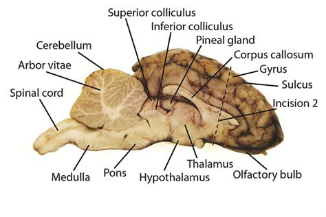
Table of Contents
A Deep Dive into the Sheep Brain Cross-Section: Anatomy, Function, and Clinical Significance
The sheep brain, a readily available and ethically sourced model organism, offers a remarkably detailed and accessible representation of mammalian brain structure. Studying a cross-section of a sheep brain provides invaluable insights into the intricate organization and functional specializations of the central nervous system. This comprehensive guide explores the key anatomical features observable in a sheep brain cross-section, their corresponding functions, and the clinical relevance of understanding this complex organ.
Gross Anatomy of the Sheep Brain Cross-Section
A midsagittal cross-section of a sheep brain reveals a stunning array of structures, broadly categorized into the cerebrum, cerebellum, and brainstem. Understanding the relationships between these regions is crucial for grasping the brain's overall functionality.
Cerebrum: The Seat of Higher Cognitive Functions
The cerebrum, the largest part of the sheep brain, dominates the cross-section. Its highly convoluted surface, characterized by gyri (ridges) and sulci (grooves), dramatically increases the surface area, accommodating vast numbers of neurons. Key features visible in a cross-section include:
-
Cerebral Cortex: The outermost layer of the cerebrum, responsible for higher-order cognitive functions like learning, memory, language, and conscious thought. Different areas of the cortex are specialized for different functions (e.g., visual cortex, auditory cortex, motor cortex). A cross-section reveals the layered structure of the cortex, with distinct cytoarchitectural differences between layers.
-
Corpus Callosum: This prominent white matter structure, clearly visible in a midsagittal section, connects the two cerebral hemispheres, facilitating interhemispheric communication and coordination. Damage to the corpus callosum can lead to a range of neurological deficits, affecting communication and coordination between the two sides of the body.
-
Lateral Ventricles: These fluid-filled cavities are part of the ventricular system, which produces and circulates cerebrospinal fluid (CSF). CSF provides cushioning and protection for the brain, removes metabolic waste products, and plays a role in maintaining a stable intracranial environment. In a cross-section, the lateral ventricles appear as C-shaped spaces within the cerebral hemispheres.
-
Basal Ganglia: Deep within the cerebrum lie the basal ganglia, a group of subcortical nuclei crucial for motor control, learning, and habit formation. These structures are not as easily visible in a gross cross-section but are crucial to understand in relation to the cerebral cortex and other brain regions. Their dysfunction is linked to movement disorders like Parkinson's disease.
Cerebellum: Master of Coordination and Balance
Located posterior to the cerebrum, the cerebellum is readily apparent in a cross-section. Its highly organized structure, characterized by numerous parallel folds (folia), is essential for motor coordination, balance, and posture.
-
Cerebellar Cortex: The outermost layer of the cerebellum, responsible for processing sensory information related to movement and coordinating motor outputs. Its highly organized structure is reflected in the parallel arrangement of its folia, which are visible even in a gross cross-section.
-
Cerebellar Nuclei: Deep within the cerebellar white matter lie the cerebellar nuclei, which relay information from the cerebellar cortex to other parts of the brain. These structures are critical for motor control and the fine-tuning of movements.
Brainstem: The Vital Connection
The brainstem, connecting the cerebrum and cerebellum to the spinal cord, is a critical structure for basic life functions. A cross-section reveals several key components:
-
Midbrain: The midbrain contains important nuclei involved in visual and auditory reflexes, as well as the substantia nigra, which plays a crucial role in movement control (and whose degeneration is linked to Parkinson's disease).
-
Pons: The pons acts as a relay station between the cerebrum and cerebellum, and also contains nuclei involved in respiration and sleep-wake cycles.
-
Medulla Oblongata: The medulla oblongata controls vital autonomic functions such as heart rate, breathing, and blood pressure. Damage to the medulla can be life-threatening.
Microscopic Anatomy and Functional Specialization
While a gross cross-section provides a general overview, microscopic examination reveals the intricate cellular architecture of the sheep brain. Different brain regions exhibit distinct cytoarchitectonic features, reflecting their specialized functions.
-
Neurons: The fundamental units of the nervous system, neurons are responsible for transmitting information throughout the brain. Their morphology varies depending on their location and function. Pyramidal neurons are characteristic of the cerebral cortex, while Purkinje cells are unique to the cerebellum.
-
Glia: Glial cells, including astrocytes, oligodendrocytes, and microglia, provide support and protection for neurons. They play critical roles in maintaining the brain's environment, regulating synaptic transmission, and responding to injury.
-
Myelin: Myelin, a fatty insulating sheath surrounding axons, speeds up nerve impulse conduction. The presence of myelin gives white matter its characteristic appearance.
Clinical Significance of Sheep Brain Cross-Section Studies
The sheep brain's similarity to the human brain makes it an invaluable model for studying neurological disorders and developing new therapies. Studying cross-sections allows for:
-
Understanding Neurological Diseases: Examining cross-sections from sheep brains affected by neurological conditions can reveal the structural and cellular changes associated with these diseases. This can provide valuable insights into disease mechanisms and potential therapeutic targets.
-
Drug Development and Testing: Sheep brains can be used to test the efficacy and safety of new drugs aimed at treating neurological disorders. Cross-sectional studies can help assess the effects of drugs on different brain regions and identify potential side effects.
-
Surgical Planning and Training: Cross-sections of sheep brains can be used for surgical planning and training. Surgeons can practice complex neurosurgical procedures on the model, improving their skills and reducing the risk of complications in human patients.
-
Comparative Neuroanatomy: Comparing cross-sections of sheep brains with those of other species allows researchers to study the evolution of brain structure and function. This can provide insights into the adaptive significance of different brain regions.
Beyond the Cross-Section: Exploring Three-Dimensional Structure
While a cross-section is extremely informative, it only provides a two-dimensional view of a three-dimensional structure. Advanced imaging techniques like MRI and CT scanning allow for a comprehensive three-dimensional visualization of the sheep brain, providing even greater detail and allowing for more sophisticated analyses.
Ethical Considerations
It's crucial to emphasize the ethical considerations surrounding the use of sheep brains for research and education. It's imperative that all procedures adhere to strict ethical guidelines and regulations, ensuring that animals are treated humanely and that their welfare is prioritized. The use of already deceased animals for educational purposes should always be done responsibly and with respect.
Conclusion
The cross-section of a sheep brain offers a unique window into the complexity and beauty of the mammalian brain. By carefully examining its anatomical features and understanding their functional significance, we can gain profound insights into the workings of the nervous system and the impact of neurological disorders. This knowledge forms the foundation for advancements in diagnosis, treatment, and prevention of brain-related diseases, highlighting the crucial role of this seemingly simple model in advancing our understanding of one of the most complex organs in the animal kingdom. The continuing study of the sheep brain, both in gross cross-section and through more advanced techniques, promises to further enhance our understanding of the brain and its functions, contributing significantly to the fields of neuroscience and medicine. The ethical and responsible use of this valuable resource remains paramount in this ongoing pursuit of knowledge.
Latest Posts
Latest Posts
-
How To Find First Term Of Arithmetic Sequence
Mar 19, 2025
-
Nursing Interventions For Patients With Schizophrenia
Mar 19, 2025
-
Three Important Parts Of Microscope Care
Mar 19, 2025
-
Lock And Key Model Of Enzyme Action
Mar 19, 2025
-
Integration Of Odd And Even Functions
Mar 19, 2025
Related Post
Thank you for visiting our website which covers about Cross Section Of A Sheep Brain . We hope the information provided has been useful to you. Feel free to contact us if you have any questions or need further assistance. See you next time and don't miss to bookmark.
