Dissection Of A Frog Labeled Parts
Muz Play
Apr 07, 2025 · 7 min read
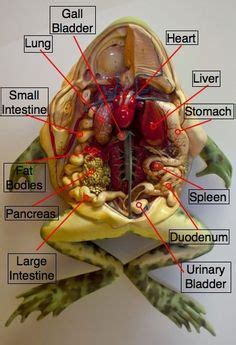
Table of Contents
Dissecting a Frog: A Comprehensive Guide to Labeled Parts
The dissection of a frog is a classic biological exercise offering invaluable hands-on experience in understanding vertebrate anatomy. This detailed guide provides a step-by-step approach to frog dissection, focusing on the identification and labeling of key anatomical structures. While this guide avoids explicit instructions for procuring a specimen (ethical considerations are paramount), it assumes you have a preserved frog ready for dissection. Remember to always follow your instructor's guidelines and prioritize safety throughout the process.
Preparing for Your Frog Dissection
Before you begin, gather the necessary materials:
- Preserved Frog: Ensure your specimen is properly preserved.
- Dissecting Tray: A sturdy tray to hold the frog and prevent spills.
- Dissecting Kit: This includes a scalpel, forceps, scissors, and probes. Sharp instruments are crucial for clean cuts.
- Gloves: Protect yourself from potential irritants and preserve the specimen.
- Paper Towels: For cleaning and absorbing excess fluid.
- Lab Coat or Apron: Protect your clothing.
- Labeled Diagram: A pre-prepared diagram to guide your identification.
- Pen or Pencil: For labeling your specimen directly (if permitted) or your diagram.
External Anatomy of the Frog: A Visual Inspection
Before making any incisions, carefully observe the external anatomy of the frog. Familiarize yourself with the following structures:
Head:
- Eyes: Large, bulging eyes protected by nictitating membranes (a transparent third eyelid).
- Tympanic Membranes (Eardrums): Located behind the eyes, these are crucial for hearing.
- Nostrils (Nares): External openings for respiration.
- Mouth: Observe the wide mouth, noting the lack of external ears.
Body:
- Skin: Smooth, moist skin that aids in respiration. Note its texture and color.
- Limbs: Examine the forelimbs (arms) and hind limbs (legs), paying attention to the number of digits (toes) on each.
- Webbing: Notice the webbing between the toes of the hind limbs, essential for swimming.
- Cloaca: The single opening at the posterior end of the body serving as the exit for the digestive, urinary, and reproductive systems.
Internal Anatomy: A Step-by-Step Dissection
Now, we'll proceed with the internal dissection. Remember to work methodically and carefully, making precise cuts.
Step 1: Initial Incisions
Using your scalpel, make a shallow incision along the midline of the frog's belly, starting just below the cloaca and extending towards the chin. Be careful not to cut too deeply and damage internal organs. You can then carefully extend this incision laterally to create a flap of skin.
Step 2: Pinning the Skin Flap
Using pins, carefully secure the skin flap to the dissecting tray. This will expose the underlying muscles and organs.
Step 3: Exposing the Muscles
Once the skin is pinned back, you will observe the underlying muscles. You'll see a thin layer of connective tissue which you can carefully cut away with your scissors or forceps to expose the muscular system. Note the location and arrangement of different muscle groups.
Step 4: Opening the Body Cavity (Coelom)
Carefully make a shallow incision through the muscle layer following the initial incision line. This will reveal the frog's coelom, the main body cavity containing the internal organs. Continue to pin back the muscle flaps to gain full access.
Step 5: Examining the Major Organs
Now you'll see several key organs. Carefully identify and examine them, referring to your labeled diagram:
- Heart: A three-chambered heart located near the anterior end of the coelom. Note its size and position.
- Lungs: Small, sac-like lungs located on either side of the heart.
- Liver: A large, dark-colored organ which is often the first organ you'll see. Note its lobes.
- Stomach: A J-shaped organ located posterior to the liver. If the frog has recently eaten, you might observe contents within.
- Small Intestine: A long, coiled tube extending from the stomach.
- Large Intestine: A shorter, wider tube leading to the cloaca.
- Spleen: A small, dark-red organ near the stomach. It plays a role in the immune system.
- Pancreas: A diffuse, light-colored gland located near the small intestine.
- Gallbladder: A small, greenish sac attached to the liver which stores bile.
- Kidneys: Bean-shaped organs located dorsally (towards the back) near the spine.
- Fat Bodies: Yellowish, fatty tissue attached to the kidneys, especially prominent in well-fed frogs. These are energy reserves.
Step 6: Exploring the Digestive System
Gently trace the path of the digestive system, from the mouth to the cloaca. Observe the transition from the esophagus to the stomach, then the small and large intestines. You may gently lift and examine different sections to see their structure.
Step 7: Examining the Circulatory System
Carefully observe the heart, noting the three chambers (two atria and one ventricle). You may carefully lift and examine the major blood vessels emerging from the heart, if your dissection allows.
Step 8: Examining the Urinary and Reproductive Systems
Locate the kidneys and observe their position relative to the other organs. The reproductive organs (testes in males, ovaries in females) are located near the kidneys and will vary significantly in size and appearance based on the frog's sex and reproductive state.
Step 9: Nervous System Observation (Optional and More Advanced)
Dissecting the nervous system requires great care and precision. If your dissection permits and you're comfortable, you can gently lift and carefully observe parts of the spinal cord. However, this is generally a more advanced stage and may be best left for more experienced students.
Labeled Diagram and Detailed Descriptions of Key Frog Anatomy
Here’s a more detailed breakdown of key anatomical structures, referencing the parts you should be able to identify during your dissection:
1. Integumentary System: The frog's skin is smooth, moist, and permeable. It plays a vital role in respiration and water absorption.
2. Muscular System: Frogs possess a complex muscular system enabling various movements, including jumping, swimming, and climbing. Observe the different muscle groups and their arrangement.
3. Skeletal System: While not explicitly dissected in a basic frog dissection, you should be aware of the underlying skeletal structure providing support and protection. Observe how the muscles are attached to bones.
4. Digestive System: The frog's digestive system is efficient at processing insects and other small prey. Trace the path of food through the esophagus, stomach, small and large intestines, and finally, the cloaca.
* **Esophagus:** Transports food from the mouth to the stomach.
* **Stomach:**  Digests food through chemical and mechanical processes.
* **Small Intestine:**  Primary site of nutrient absorption.
* **Large Intestine:**  Absorbs water and forms waste.
* **Cloaca:** The common exit for the digestive, urinary, and reproductive systems.
5. Respiratory System: Frogs use both lungs and their skin for gas exchange. Examine the small lungs.
6. Circulatory System: Frogs have a three-chambered heart (two atria and one ventricle), a less efficient system compared to mammals. Note the position of the heart and major blood vessels (if possible and safe to dissect).
7. Urinary System: The kidneys filter waste products from the blood, producing urine that is expelled through the cloaca.
8. Nervous System: The brain, spinal cord, and peripheral nerves control the frog's actions and responses. Observe with extreme caution if allowed as part of the dissection.
9. Reproductive System: The reproductive organs (testes in males, ovaries in females) produce gametes (sperm and eggs) for reproduction. The location and size vary considerably between sexes and reproductive stages.
10. Endocrine System: While not directly visible in a basic dissection, understand the endocrine system's role in hormone production and regulation.
Post-Dissection Cleanup and Ethical Considerations
After completing your dissection, properly dispose of the frog and all materials according to your instructor’s instructions. Dispose of sharp instruments carefully. Clean your dissecting tray and tools thoroughly. Respect the life of the animal, even in its preserved state, and remember the importance of ethical practices in scientific research.
Conclusion: Learning from Frog Dissection
Frog dissection is a powerful learning tool. It provides invaluable insights into vertebrate anatomy, fostering a deeper understanding of biological principles. While challenging, the careful and respectful examination of a frog offers a unique opportunity for hands-on learning and a more comprehensive understanding of life sciences. Remember to always prioritize safety and ethical considerations throughout the process. By carefully following this guide, you'll be able to confidently identify and label the key anatomical structures of a frog.
Latest Posts
Latest Posts
-
Which One Of The Following Is A Chemical Change
Apr 10, 2025
-
Write A Function In Standard Form
Apr 10, 2025
-
Row Vs Column Percentages Independent Variable
Apr 10, 2025
-
Which Molecule Is Expected To Have The Smallest Pka
Apr 10, 2025
-
How Many Electrons Are Shared In A Double Covalent Bond
Apr 10, 2025
Related Post
Thank you for visiting our website which covers about Dissection Of A Frog Labeled Parts . We hope the information provided has been useful to you. Feel free to contact us if you have any questions or need further assistance. See you next time and don't miss to bookmark.