During Contraction Of A Muscle Calcium Ions Bind To The
Muz Play
Mar 24, 2025 · 6 min read
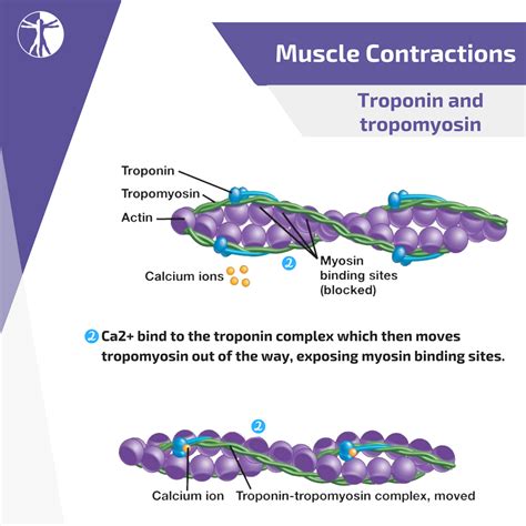
Table of Contents
During Contraction of a Muscle, Calcium Ions Bind To The… Troponin Complex: A Deep Dive into Muscle Contraction
Muscle contraction, a fundamental process enabling movement, is a complex interplay of biochemical events. At the heart of this process lies the interaction between calcium ions (Ca²⁺) and the troponin complex within muscle fibers. This article will delve deep into the mechanisms of muscle contraction, focusing specifically on the crucial role of calcium ions binding to troponin and the subsequent events leading to muscle shortening. We'll explore the intricacies of this process, considering different muscle types and the implications of disruptions to this finely tuned system.
The Players: Actin, Myosin, and the Troponin Complex
Before we explore the calcium-troponin interaction, let's briefly review the key players in muscle contraction:
Actin Filaments: The Thin Filaments
Actin filaments are thin protein filaments comprising primarily actin monomers arranged in a double helix. These filaments are integral to the sarcomere, the basic contractile unit of a muscle fiber. They contain binding sites for myosin heads, crucial for force generation. However, these binding sites are normally blocked in a resting muscle.
Myosin Filaments: The Thick Filaments
Myosin filaments are thicker filaments composed of numerous myosin molecules. Each myosin molecule has a head and a tail. The myosin heads possess ATPase activity, enabling them to bind to actin, hydrolyze ATP, and generate force. These heads act as molecular motors, driving the sliding filament mechanism of muscle contraction.
The Troponin Complex: The Calcium Sensor
The troponin complex, nestled within the actin filament, acts as a crucial calcium sensor. It's composed of three subunits:
- Troponin C (TnC): This subunit has a high affinity for calcium ions. Binding of calcium to TnC is the pivotal event initiating muscle contraction.
- Troponin I (TnI): This subunit inhibits the interaction between actin and myosin in the absence of calcium. It binds to both actin and troponin T.
- Troponin T (TnT): This subunit anchors the troponin complex to tropomyosin.
Tropomyosin: The Regulator
Tropomyosin is a filamentous protein that winds along the actin filament, covering the myosin-binding sites on actin. In the absence of calcium, tropomyosin effectively blocks these sites, preventing myosin-actin interaction and muscle contraction.
The Calcium-Troponin Interaction: Initiating Muscle Contraction
The process begins with the arrival of a nerve impulse at the neuromuscular junction. This impulse triggers the release of acetylcholine, a neurotransmitter, which subsequently depolarizes the muscle fiber membrane. This depolarization spreads into the transverse tubules (T-tubules) of the muscle fiber, leading to the release of calcium ions from the sarcoplasmic reticulum (SR), a specialized intracellular calcium store.
The released calcium ions diffuse into the cytoplasm of the muscle fiber and bind to troponin C (TnC). This binding causes a conformational change in TnC, altering the shape of the entire troponin complex. This conformational change is the critical step.
The crucial consequence of calcium binding to TnC is the movement of tropomyosin. The conformational shift in the troponin complex pulls tropomyosin away from the myosin-binding sites on the actin filament. This uncovers the binding sites, making them accessible to the myosin heads.
The Cross-Bridge Cycle: Generating Force
Once the myosin-binding sites are exposed, the cross-bridge cycle begins:
- Cross-bridge formation: A myosin head, carrying ADP and inorganic phosphate (Pi), binds to an exposed myosin-binding site on actin.
- Power stroke: The myosin head undergoes a conformational change, releasing ADP and Pi. This change pivots the myosin head, generating force and pulling the actin filament towards the center of the sarcomere.
- Cross-bridge detachment: An ATP molecule binds to the myosin head, causing it to detach from the actin filament.
- ATP hydrolysis: The ATP molecule is hydrolyzed to ADP and Pi, resetting the myosin head to its high-energy conformation, ready for another cycle.
This cycle repeats multiple times, resulting in the sliding of actin filaments over myosin filaments, shortening the sarcomere and ultimately causing muscle contraction. The process continues as long as calcium ions remain bound to troponin C and ATP is available.
Relaxation: The Role of Calcium Removal
Muscle relaxation occurs when the nerve impulse ceases. This leads to the cessation of acetylcholine release and the subsequent re-establishment of the muscle fiber membrane potential. The sarcoplasmic reticulum then actively pumps calcium ions back into its lumen using calcium ATPase pumps.
As cytoplasmic calcium levels fall, calcium ions detach from troponin C. This causes a reversal of the conformational changes in the troponin complex, resulting in tropomyosin moving back to its original position, blocking the myosin-binding sites on actin. This prevents further cross-bridge cycling and causes muscle relaxation.
Variations in Muscle Contraction: Different Muscle Types
While the fundamental mechanism of calcium-troponin interaction remains the same, there are variations in the speed and duration of contraction among different muscle types:
Skeletal Muscle: Fast and Powerful Contractions
Skeletal muscles are responsible for voluntary movements. They exhibit rapid and powerful contractions, characterized by a rapid release and uptake of calcium ions by the sarcoplasmic reticulum. The speed and efficiency of this calcium handling are crucial for their function.
Cardiac Muscle: Rhythmic and Sustained Contractions
Cardiac muscle cells exhibit rhythmic and sustained contractions, responsible for the pumping action of the heart. The calcium handling in cardiac muscle is more complex, involving both intracellular and extracellular calcium sources. The interplay between calcium influx from the extracellular space and the release of calcium from the SR contributes to the prolonged contraction of cardiac muscle.
Smooth Muscle: Slow and Sustained Contractions
Smooth muscles are found in the walls of internal organs and blood vessels. They exhibit slow and sustained contractions, often regulated by hormonal and neural signals. The calcium-handling mechanisms in smooth muscle are unique, often involving calcium influx through various membrane channels, and the interaction of calcium with calmodulin, another calcium-binding protein.
Clinical Significance: Disorders of Muscle Contraction
Disruptions to the calcium-troponin interaction can lead to various muscle disorders. These disruptions can stem from:
- Genetic mutations: Mutations affecting the genes encoding troponin proteins can cause familial hypertrophic cardiomyopathy, a condition characterized by thickening of the heart muscle.
- Metabolic disorders: Conditions like hypocalcemia (low blood calcium levels) can impair muscle contraction, leading to muscle weakness and cramps.
- Neurological disorders: Disorders affecting nerve impulses can disrupt the release of acetylcholine at the neuromuscular junction, leading to muscle weakness or paralysis.
Understanding the intricacies of calcium-troponin interaction is crucial for diagnosing and treating various muscle disorders.
Conclusion: A Precisely Regulated Process
The binding of calcium ions to troponin C is a pivotal event triggering muscle contraction. This exquisitely regulated process involves a complex interplay of proteins and ions, working in concert to generate force and movement. Understanding this process, including its variations across muscle types and the consequences of disruptions, is essential for comprehending the physiological basis of movement and for advancing the diagnosis and treatment of muscle-related disorders. Future research focusing on the molecular details of calcium handling and its regulation will continue to shed light on this crucial biological process. The study of muscle contraction is a dynamic field, continually revealing new insights into this fundamental biological mechanism, with implications spanning various areas of biology and medicine. Further exploration into the intricacies of this system promises continued advancements in our understanding of health and disease.
Latest Posts
Latest Posts
-
What Is The Angle Of Reflection
Mar 30, 2025
-
As A Balloon Is Inflated What Happens To The Pressure
Mar 30, 2025
-
How To Find The Heat Capacity Of A Calorimeter
Mar 30, 2025
-
Define The Simplest Form Of A Rate
Mar 30, 2025
-
How To Add Radicals With Different Radicands
Mar 30, 2025
Related Post
Thank you for visiting our website which covers about During Contraction Of A Muscle Calcium Ions Bind To The . We hope the information provided has been useful to you. Feel free to contact us if you have any questions or need further assistance. See you next time and don't miss to bookmark.
