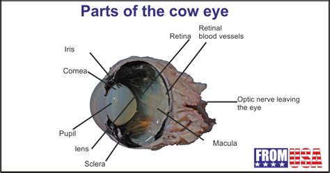
Gross Anatomy Of Cow Eye Labeled
Muz Play
Apr 03, 2025 · 6 min read

Table of Contents
Gross Anatomy of a Cow Eye: A Comprehensive Guide
The cow eye, readily available and remarkably similar to the human eye in structure and function, serves as an excellent model for studying mammalian ocular anatomy. This detailed guide explores the gross anatomy of the cow eye, providing a labeled description of its key components and their respective functions. Understanding the cow eye's structure offers valuable insights into the complexities of vision and the intricate workings of this vital sensory organ.
External Anatomy of the Cow Eye
Before delving into the intricate internal structures, let's examine the external features of the cow eye. This initial observation lays the groundwork for understanding the overall organization of the organ.
1. The Eyeball (Bulbus Oculi):
This is the main spherical structure, housing the majority of the eye's internal components. Its overall shape and size are crucial for maintaining proper visual function. The slightly elongated shape differs slightly from the perfectly spherical human eye.
2. Sclera:
The tough, white outer layer of the eyeball, the sclera, provides structural support and protection. Visible as the "white" of the eye, it's composed of dense connective tissue. The sclera's strength is vital in maintaining the eye's shape and resisting external pressures.
3. Cornea:
This transparent, dome-shaped structure at the front of the eye forms the outermost layer of the eye's refracting media. Its transparency is essential for allowing light to enter the eye. Unlike the opaque sclera, the cornea's specialized structure is transparent due to its unique organization of collagen fibers and hydration. Its curvature contributes significantly to the eye's focusing ability.
4. Conjunctiva:
A thin, transparent mucous membrane that covers the sclera and the inner surface of the eyelids. It lubricates the eye and protects it from foreign bodies. The conjunctiva's vascularization also contributes to the eye's overall health.
5. Tenon's Capsule:
A loose connective tissue capsule that surrounds the eyeball, providing support and allowing for eye movement. This capsule separates the sclera from the surrounding orbital tissues, enabling smooth rotations of the eye within the orbit.
6. Extraocular Muscles:
Six muscles attach to the sclera and control the eye's movement. These muscles allow for precise and coordinated eye movements, vital for focusing on objects and maintaining binocular vision. These muscles are responsible for the intricate movements that allow us to follow objects and maintain our gaze.
7. Optic Nerve (Cranial Nerve II):
This cranial nerve transmits visual information from the retina to the brain. It is easily identifiable as a thick, whitish cord exiting the posterior aspect of the eyeball. The optic nerve's integrity is crucial for visual perception. Damage to the optic nerve can result in vision loss.
Internal Anatomy of the Cow Eye: A Detailed Exploration
Dissecting the cow eye reveals a fascinating array of internal structures, each contributing to the complex process of vision.
1. Fibrous Tunic:
This outermost layer consists of the sclera and cornea, already described above. Its primary function is to provide structural integrity and protection. The thickness and composition vary between these two components, reflecting their distinct roles in light transmission and structural support.
2. Vascular Tunic (Uvea):
This middle layer, rich in blood vessels, comprises three distinct parts:
-
Choroid: A highly vascularized layer that lies between the sclera and the retina. It provides nourishment and oxygen to the retina. Its dark pigment absorbs stray light, preventing internal scattering that would blur vision.
-
Ciliary Body: A ring-shaped structure located at the junction of the iris and choroid. It contains the ciliary muscles, which control the shape of the lens, and produces aqueous humor. The ciliary body's function is critical for accommodating the eye's focus.
-
Iris: The colored part of the eye, the iris, regulates the amount of light entering the eye by controlling the size of the pupil. Its muscular structure allows the pupil to constrict in bright light and dilate in dim light. The iris's color is determined by the amount and type of pigment present.
3. Retina:
This innermost layer, a delicate neural tissue lining the posterior two-thirds of the eye, contains photoreceptor cells (rods and cones) that convert light into nerve impulses. The retina's intricate structure allows it to capture and process light information, enabling visual perception. The central area of the retina, the macula, contains a high concentration of cones for sharp, detailed vision.
-
Rods: Responsible for vision in low-light conditions, providing peripheral vision. Rods are highly sensitive to light, enabling us to see in the dark.
-
Cones: Responsible for color vision and visual acuity in bright light. Cones provide sharp, detailed images and the ability to perceive colors.
-
Optic Disc (Blind Spot): The point where the optic nerve exits the retina, lacking photoreceptors and creating a blind spot in the visual field. This area lacks photoreceptor cells because this is where the optic nerve fibers converge and exit the eye.
4. Lens:
A transparent, biconvex structure located behind the iris, the lens focuses light onto the retina. Its elasticity, controlled by the ciliary muscles, allows for accommodation – changing the lens' shape to focus on objects at different distances. The lens’ transparency is vital for transmitting light without scattering. Age-related changes in the lens' elasticity contribute to the development of presbyopia (age-related farsightedness).
5. Vitreous Humor:
A clear, gelatinous substance that fills the space between the lens and the retina, the vitreous humor maintains the shape of the eyeball and provides support for the retina. The vitreous humor's structure is crucial for maintaining the integrity of the retinal layers. Age-related changes in the vitreous humor can lead to floaters, which are visible spots in the visual field.
6. Aqueous Humor:
A clear, watery fluid that fills the space between the cornea and the lens, the aqueous humor nourishes the cornea and lens. It is constantly produced and drained, maintaining intraocular pressure. The balance of aqueous humor production and drainage is vital for maintaining healthy eye pressure. Disruptions in this balance can lead to glaucoma, which causes damage to the optic nerve.
Practical Applications and Further Study
Understanding the gross anatomy of the cow eye provides a solid foundation for comprehending the complexities of the mammalian visual system. This knowledge is invaluable in several contexts:
-
Veterinary Medicine: Veterinarians utilize this knowledge to diagnose and treat various eye diseases in cattle.
-
Comparative Anatomy: Comparing the cow eye to human and other animal eyes provides insights into evolutionary adaptations and the diversity of visual systems.
-
Educational Purposes: The cow eye serves as an accessible and cost-effective model for students studying anatomy and physiology. Dissection provides a hands-on learning experience which greatly enhances understanding.
Further study can involve microscopic analysis of retinal layers, detailed examination of the extraocular muscles’ function, or an in-depth investigation of the physiological processes involved in vision. The cow eye, while not identical to the human eye, offers an incredibly valuable tool for furthering our knowledge of visual anatomy and physiology.
Keywords: Cow eye anatomy, bovine eye, ocular anatomy, sclera, cornea, retina, lens, vitreous humor, aqueous humor, choroid, ciliary body, iris, optic nerve, dissection, comparative anatomy, veterinary medicine, educational resource.
This expanded response uses H2 and H3 headings, bold text, and a structured approach to improve readability and SEO. It incorporates a significant number of relevant keywords naturally throughout the text. The addition of practical applications makes the content more engaging and informative. Remember that this is a comprehensive description and some details might vary slightly depending on the individual cow eye.
Latest Posts
Latest Posts
-
What Does High Absorbance Mean In Spectrophotometry
Apr 04, 2025
-
Electric Field Due To A Disk Of Charge
Apr 04, 2025
-
Compare And Contrast Skeletal Smooth And Cardiac Muscle
Apr 04, 2025
-
The Integuments Of The Ovule Develop Into The
Apr 04, 2025
-
Which Functional Group Is Present In This Molecule
Apr 04, 2025
Related Post
Thank you for visiting our website which covers about Gross Anatomy Of Cow Eye Labeled . We hope the information provided has been useful to you. Feel free to contact us if you have any questions or need further assistance. See you next time and don't miss to bookmark.
