How To Tell Left And Right Ulna
Muz Play
Apr 02, 2025 · 5 min read

Table of Contents
How to Tell Left and Right Ulna: A Comprehensive Guide for Anatomy Students and Professionals
Identifying left and right ulna bones can be tricky, even for seasoned anatomists. This comprehensive guide will equip you with the knowledge and techniques to confidently differentiate between a left and a right ulna, regardless of the viewing angle or preparation state. We'll explore various anatomical features, practical tips, and helpful imagery to ensure you master this crucial skill.
Understanding the Ulna's Anatomy: The Foundation of Identification
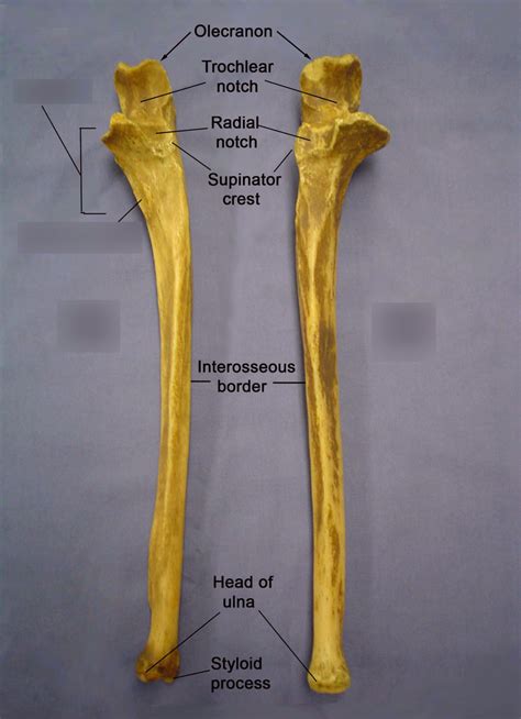
Before diving into identification techniques, let's establish a solid understanding of the ulna's key anatomical features. The ulna, one of the two bones in the forearm (the other being the radius), is a long bone with a unique shape that dictates its left-right orientation. Key features include:
1. The Trochlear Notch: The Hinge of the Elbow
The trochlear notch is a large, C-shaped concave surface located at the proximal end of the ulna. This notch articulates with the trochlea of the humerus, forming the hinge joint of the elbow. Critically, the orientation of this notch is crucial for determining left and right.
2. The Coronoid Process: A Crucial Identifying Landmark
The coronoid process projects anteriorly from the proximal end of the ulna, just below the trochlear notch. Its shape and position relative to the trochlear notch are significant identification clues.
3. The Olecranon Process: The Point of the Elbow
The olecranon process is the prominent posterior projection at the proximal end of the ulna. It forms the bony prominence of your elbow. While not uniquely defining left or right, its position relative to the trochlear notch is important in the overall assessment.
4. The Radial Notch: Articulation with the Radius
Located on the lateral side of the proximal ulna, the radial notch is a small, concave articular surface that receives the head of the radius. Its position, always on the lateral aspect, helps confirm orientation, especially when viewed from the proximal end.
5. The Ulnar Tuberosity: Muscle Attachment Site
The ulnar tuberosity is a roughened projection located on the anterior surface of the ulna, just distal to the coronoid process. This serves as an attachment point for the brachialis muscle. While not directly related to left-right identification, it can be a useful secondary confirmation point.
6. The Styloid Process: Distal Landmark
At the distal end of the ulna, the styloid process is a pointed projection. Its position helps to confirm the overall orientation and can be useful as a secondary marker when viewed from the distal aspect.
Techniques for Identifying Left and Right Ulna
Now that we've established the key anatomical landmarks, let's explore practical techniques for confidently identifying the left and right ulna.
1. The Proximal End Approach: Focusing on the Trochlear Notch
This is arguably the most reliable method. Hold the bone with the proximal end facing you. Imagine the trochlear notch as a horseshoe. The orientation of the horseshoe is key:
- Left Ulna: If you were to place your thumb into the "horseshoe" of the trochlear notch, your thumb would point towards the ulnar tuberosity. The coronoid process would be on the side closer to your fingers.
- Right Ulna: The "horseshoe" of the trochlear notch would be oriented such that your thumb, placed into the notch, would point away from the ulnar tuberosity. The coronoid process would be closer to your palm.
Important Note: Practice visualizing this “horseshoe” analogy. Repeated practice with physical specimens or anatomical models is crucial for mastery.
2. The Distal End Approach: Using the Styloid Process and Orientation
While less definitive than the proximal approach, the distal end can provide supporting evidence. Hold the distal end of the bone:
- Left Ulna: The styloid process will point medially (towards the midline of the body).
- Right Ulna: The styloid process will point medially, too. But importantly, when positioned with the styloid process towards the body midline, its orientation is such that the distal articulation surface points towards the radial side.
3. The Articulatory Surface Approach: Considering the Radius
Consider the articular surfaces of the ulna. The radial notch, always positioned laterally, will help you understand where the radius should articulate. This articulation adds an extra level of confirmation to your left-right assessment. This method works effectively in conjunction with other methods.
4. The "Holding the Bone" Method: Mimicking Anatomical Position
Hold the bone as you would if it were in its anatomical position in the body. Your left hand mimics the left ulna, and your right hand mimics the right ulna. This kinesthetic approach can strengthen your understanding of anatomical position.
Common Mistakes and Troubleshooting
Even with the techniques above, there's always a chance of error. Be aware of these common mistakes:
- Rotation: Misinterpreting the orientation due to rotation of the bone. Ensure the bone is in a stable, easily observable position before attempting identification.
- Incomplete Observation: Failing to consider all relevant landmarks. Carefully examine the proximal and distal ends, and articular surfaces.
- Lack of Practice: Insufficient hands-on experience with real or model specimens. Consistent practice is crucial for developing the necessary visual and tactile skills.
Advanced Techniques and Considerations
For more advanced learners, these techniques can further enhance your identification skills:
- Radiographic Analysis: X-rays can be invaluable, particularly in the context of fracture analysis. Understanding the characteristic radiographic appearances of left and right ulnae, especially considering the proximal and distal articular surfaces is important.
- Comparative Anatomy: Comparing the ulna to the radius and other forearm bones will refine your understanding of its unique features and its position within the skeletal framework.
Conclusion: Mastering Ulna Identification
Mastering the ability to differentiate between left and right ulnae is a crucial skill for anatomy students, medical professionals, and anyone working with human skeletal remains or anatomical models. By utilizing the techniques outlined in this guide, combining multiple identification methods, and incorporating regular practice, you can build your confidence and develop a robust ability to confidently determine the laterality of the ulna. Remember that consistent practice and a holistic approach incorporating multiple anatomical landmarks are key to successfully identifying left and right ulnae. Develop your spatial reasoning skills and always double check your identification using multiple methods. The more you practice, the more intuitive this process will become.
Latest Posts
Latest Posts
-
Difference Between A Somatic Cell And A Gamete
Apr 03, 2025
-
Match The Structure Process To The Letter
Apr 03, 2025
-
Find The Basis Of The Subspace
Apr 03, 2025
-
Why Is Immersion Oil Used With The 100x Objective
Apr 03, 2025
-
What Is Shared In A Covalent Bond
Apr 03, 2025
Related Post
Thank you for visiting our website which covers about How To Tell Left And Right Ulna . We hope the information provided has been useful to you. Feel free to contact us if you have any questions or need further assistance. See you next time and don't miss to bookmark.
