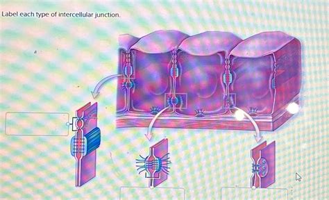
Label Each Type Of Intercellular Junction
Muz Play
Apr 02, 2025 · 6 min read

Table of Contents
Labeling Each Type of Intercellular Junction: A Comprehensive Guide
Intercellular junctions are specialized structures that hold cells together and facilitate communication between them. These junctions are crucial for maintaining tissue integrity, coordinating cellular activities, and ensuring the proper functioning of organs and systems within the body. Understanding the different types of intercellular junctions and their specific roles is fundamental to comprehending the complexities of multicellular organisms. This comprehensive guide will delve into the various types of intercellular junctions, detailing their structures, functions, and locations within the body.
The Major Families of Intercellular Junctions
Intercellular junctions are broadly categorized into several families based on their structure and primary function. These families include:
- Tight Junctions (Zonula Occludens): These junctions form a continuous seal around the apical region of epithelial cells, preventing the passage of molecules between cells.
- Adherens Junctions (Zonula Adherens): These junctions provide strong adhesion between cells, contributing to the structural integrity of tissues.
- Desmosomes (Macula Adherens): These junctions provide spot-like adhesion between cells, offering robust mechanical strength to tissues subjected to stress.
- Gap Junctions (Nexus): These junctions facilitate direct communication between cells through the exchange of ions and small molecules.
- Hemidesmosomes: These junctions anchor epithelial cells to the underlying basement membrane.
Detailed Examination of Each Junction Type
1. Tight Junctions (Zonula Occludens)
Structure: Tight junctions are characterized by the fusion of the outer leaflets of the plasma membranes of adjacent cells. Transmembrane proteins, primarily claudins and occludins, form strands that interact across the intercellular space, creating a tight seal.
Function: The primary function of tight junctions is to regulate paracellular transport—the movement of substances between cells. They form a selective barrier, preventing the passage of ions, water, and larger molecules while allowing the passage of certain specific molecules. This function is crucial in maintaining the integrity of epithelial barriers, such as the intestinal lining and the blood-brain barrier. The degree of tightness varies across different tissues, reflecting the specific functional requirements of that tissue. For example, tight junctions in the intestine are highly impermeable to prevent the passage of harmful substances, while those in the kidney are more permeable to allow for selective reabsorption of molecules.
Location: Tight junctions are found in epithelial tissues throughout the body, including the lining of the digestive tract, the kidneys, the blood-brain barrier, and the blood-testis barrier.
2. Adherens Junctions (Zonula Adherens)
Structure: Adherens junctions are characterized by the presence of cadherin transmembrane proteins. These cadherins bind to each other in the intercellular space, connecting the actin cytoskeletons of adjacent cells. The intracellular domain of cadherins interacts with catenins, which link the cadherins to the actin filaments.
Function: Adherens junctions play a critical role in cell-cell adhesion, providing structural support and maintaining tissue integrity. They contribute to the formation of epithelial sheets and are involved in various cellular processes, including cell migration and morphogenesis. The strong adhesion provided by these junctions is crucial for resisting mechanical stress. Furthermore, their connection to the actin cytoskeleton allows for coordinated cell behavior within a tissue.
Location: Adherens junctions are found in epithelial tissues, as well as in certain types of muscle tissue. They are often located just below tight junctions in epithelial cells.
3. Desmosomes (Macula Adherens)
Structure: Desmosomes are spot-like junctions that provide strong adhesion between cells. They are characterized by the presence of cadherins (desmogleins and desmocollins), which interact in the intercellular space. The intracellular domains of these cadherins are linked to intermediate filaments, primarily keratin, within the cytoplasm. This connection to the intermediate filament network provides robust mechanical strength to the cell-cell junction.
Function: The primary function of desmosomes is to provide strong mechanical adhesion between cells, particularly in tissues subjected to high levels of shear stress or tensile force. Their role is crucial in maintaining the structural integrity of tissues such as the epidermis (outer layer of skin), the heart muscle, and the uterine cervix.
Location: Desmosomes are abundant in tissues that experience significant mechanical stress, including the epidermis, cardiac muscle, and uterine cervix.
4. Gap Junctions (Nexus)
Structure: Gap junctions are specialized intercellular channels that facilitate direct communication between cells. They are formed by connexin proteins, which assemble to create connexons. Six connexin molecules form a single connexon, and two connexons from adjacent cells align to create a channel that spans the intercellular space. These channels allow for the passage of small molecules and ions, such as calcium and cyclic AMP.
Function: Gap junctions allow for rapid cell-to-cell communication, enabling coordinated activity within tissues. This communication is crucial for various physiological processes, including heart muscle contraction, nerve impulse transmission, and embryonic development. The exchange of ions and small molecules through gap junctions allows for the synchronized activity of cells, ensuring efficient tissue function. The ability to open and close these channels also allows for regulation of intercellular communication, responding to various stimuli.
Location: Gap junctions are found in a variety of tissues, including heart muscle, smooth muscle, neurons, and epithelial cells.
5. Hemidesmosomes
Structure: Hemidesmosomes are structurally similar to desmosomes but serve a different function. They anchor epithelial cells to the underlying basement membrane, a specialized extracellular matrix. Instead of cadherins, they utilize integrins, transmembrane proteins that bind to components of the extracellular matrix such as laminin and collagen. The intracellular domain of integrins connects to intermediate filaments within the cell.
Function: The primary function of hemidesmosomes is to provide strong adhesion between epithelial cells and the basement membrane. This anchoring is essential for maintaining the integrity of epithelial layers and providing stability to tissues. They play a critical role in preventing tissue detachment and maintaining the structural integrity of the epithelium in response to mechanical stress.
Location: Hemidesmosomes are found at the basal surface of epithelial cells, anchoring them to the basement membrane.
Clinical Significance of Intercellular Junctions
Dysfunction of intercellular junctions is implicated in a variety of diseases. For instance:
- Pemphigus: This autoimmune disease targets desmosomal proteins, leading to blistering of the skin and mucous membranes.
- Certain cancers: Alterations in cell-cell adhesion mediated by adherens junctions and desmosomes can promote cancer cell invasion and metastasis.
- Inflammatory bowel disease: Disrupted tight junctions in the intestinal epithelium contribute to increased intestinal permeability and inflammation.
Conclusion
Intercellular junctions are essential structures that contribute to the integrity, function, and communication within tissues. The distinct types of junctions—tight junctions, adherens junctions, desmosomes, gap junctions, and hemidesmosomes—each play unique and vital roles in maintaining tissue homeostasis and coordinating cellular activities. A comprehensive understanding of these junctions is paramount in various fields, from basic biology to medicine, offering valuable insights into the intricacies of multicellular life and the pathophysiology of various diseases. Further research continues to unveil the precise molecular mechanisms governing the formation, function, and regulation of these critical structures.
Latest Posts
Latest Posts
-
What Is The Most Complex Level Of Organization
Apr 03, 2025
-
What Determines The Volume Of Gas
Apr 03, 2025
-
Non Mendelian Genetics Practice Packet Answers
Apr 03, 2025
-
Similarities Between Endocrine And Nervous System
Apr 03, 2025
-
How To Choose U And Dv In Integration By Parts
Apr 03, 2025
Related Post
Thank you for visiting our website which covers about Label Each Type Of Intercellular Junction . We hope the information provided has been useful to you. Feel free to contact us if you have any questions or need further assistance. See you next time and don't miss to bookmark.
