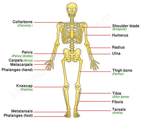
Label The Bones Of The Appendicular Skeleton
Muz Play
Apr 06, 2025 · 7 min read

Table of Contents
Label the Bones of the Appendicular Skeleton: A Comprehensive Guide
The appendicular skeleton, a fascinating and complex system, forms the appendages of our bodies – the limbs that allow us to move, grasp, and interact with the world. Understanding its intricate structure is crucial for anyone studying anatomy, physiology, or related fields. This comprehensive guide will take you on a journey through the bones of the appendicular skeleton, explaining their location, function, and key features. We'll cover both the upper and lower limbs, providing you with a detailed, easily understandable roadmap to mastering this essential anatomical knowledge.
The Upper Limb: A Detailed Exploration
The upper limb, responsible for fine motor skills and manipulation, consists of several key regions: the shoulder girdle, the arm, the forearm, and the hand. Let's examine each in detail.
The Shoulder Girdle: The Foundation of Movement
The shoulder girdle, also known as the pectoral girdle, provides the connection between the upper limb and the axial skeleton. It consists of two bones:
-
Clavicle (Collarbone): This elongated, S-shaped bone is easily palpable just beneath the skin. It articulates medially with the sternum (breastbone) and laterally with the scapula (shoulder blade), forming the sternoclavicular and acromioclavicular joints respectively. The clavicle plays a crucial role in stabilizing the shoulder joint and transmitting forces from the upper limb to the axial skeleton. Remember: fractures of the clavicle are common injuries, often resulting from falls or direct trauma.
-
Scapula (Shoulder Blade): A large, flat, triangular bone situated on the posterior aspect of the thorax. It possesses several important features: the acromion (a bony projection that articulates with the clavicle), the coracoid process (a hook-like structure providing attachment points for muscles), the glenoid cavity (a shallow socket that articulates with the head of the humerus), and the spine of the scapula (a prominent ridge running across the posterior surface). The scapula’s unique structure allows for a wide range of motion at the shoulder joint. Key takeaway: understanding the scapula's anatomy is crucial for comprehending the biomechanics of shoulder movements.
The Arm: The Humerus
The arm contains only one bone:
- Humerus: The longest and largest bone of the upper limb. This long bone has a proximal head that articulates with the glenoid cavity of the scapula, forming the glenohumeral joint (shoulder joint). The humerus also possesses several prominent features: the greater and lesser tubercles (sites for muscle attachment), the deltoid tuberosity (a roughened area for the deltoid muscle attachment), the medial and lateral epicondyles (points of attachment for forearm muscles), and the trochlea and capitulum (articulations with the forearm bones). Important note: The humerus is susceptible to fractures, particularly at the surgical neck (a common site for fractures in older adults) and the shaft.
The Forearm: Radius and Ulna
The forearm comprises two bones:
-
Radius: Situated laterally (on the thumb side) of the forearm. The proximal end of the radius articulates with the capitulum of the humerus and the ulna, forming the elbow joint. The distal end articulates with the carpal bones of the wrist. Remember: the radius is crucial for pronation and supination (rotation of the forearm).
-
Ulna: Located medially (on the pinky finger side) of the forearm. The proximal end forms the olecranon process (the bony point of the elbow) which articulates with the trochlea of the humerus. The distal end articulates with the radius and the carpal bones. Key point: The ulna is less involved in movements of the hand compared to the radius.
The Hand: Carpals, Metacarpals, and Phalanges
The hand, a marvel of intricate engineering, is divided into three main regions:
-
Carpals (Wrist Bones): Eight small, irregularly shaped bones arranged in two rows. These bones are crucial for wrist flexibility and stability. Remember to learn the names and arrangement of each carpal bone: scaphoid, lunate, triquetrum, pisiform, trapezium, trapezoid, capitate, and hamate. Their specific arrangement is vital for complex wrist movements.
-
Metacarpals (Palm Bones): Five long bones, numbered I-V from the thumb to the pinky finger. These bones articulate proximally with the carpals and distally with the phalanges. Key Feature: The metacarpals form the framework of the palm.
-
Phalanges (Finger Bones): Fourteen bones making up the fingers. The thumb possesses two phalanges (proximal and distal), while the other fingers have three (proximal, middle, and distal). Important detail: The precise arrangement and articulation of the phalanges allow for the dexterity and fine motor skills of the hand.
The Lower Limb: Supporting Our Weight and Enabling Locomotion
The lower limb, responsible for supporting our body weight and enabling locomotion, comprises several key regions: the pelvic girdle, the thigh, the leg, and the foot.
The Pelvic Girdle: Strong and Stable
Unlike the shoulder girdle, the pelvic girdle is a more robust structure designed to support the weight of the upper body and transmit forces to the lower limbs. It is formed by two hip bones (ossa coxae), the sacrum, and the coccyx.
-
Hip Bones (Ossa Coxae): Each hip bone is formed by the fusion of three bones: the ilium, ischium, and pubis. These bones meet at the acetabulum, a deep socket that articulates with the head of the femur. Important note: The hip bones are large and strong, providing a stable base for the lower limb.
-
Sacrum: A triangular bone formed by the fusion of five sacral vertebrae. It articulates with the ilium of the hip bone, forming the sacroiliac joint. Key point: The sacrum contributes significantly to the stability of the pelvic girdle.
-
Coccyx: A small, triangular bone formed by the fusion of four rudimentary vertebrae. It provides little structural support but plays a role in muscle attachment.
The Thigh: The Femur
The thigh contains only one bone:
- Femur (Thigh Bone): The longest and strongest bone in the human body. The proximal end possesses a head that articulates with the acetabulum of the hip bone, forming the hip joint. The distal end articulates with the tibia and patella. Remember: the femur's robust structure is essential for weight-bearing and locomotion. Several prominent features include the greater and lesser trochanters (sites of muscle attachment) and the condyles (articulations with the tibia).
The Leg: Tibia and Fibula
The leg consists of two bones:
-
Tibia (Shin Bone): The weight-bearing bone of the leg. It is located medially and articulates proximally with the femur and distally with the talus (ankle bone). Key Feature: The tibia is thicker and stronger than the fibula.
-
Fibula: A slender bone located laterally in the leg. It articulates proximally with the tibia and distally with the talus. It contributes to ankle stability and provides attachment points for muscles. Important note: The fibula is less involved in weight-bearing compared to the tibia.
The Foot: Tarsals, Metatarsals, and Phalanges
The foot, like the hand, is a complex structure designed for weight-bearing, balance, and locomotion. It is divided into three regions:
-
Tarsals (Ankle Bones): Seven bones forming the posterior part of the foot. The largest tarsal is the calcaneus (heel bone). The talus articulates with the tibia and fibula, forming the ankle joint. Remember: Understanding the arrangement of the tarsal bones is essential for comprehending foot biomechanics.
-
Metatarsals (Foot Bones): Five long bones that form the middle part of the foot. They articulate proximally with the tarsals and distally with the phalanges. Key feature: These bones contribute to the arch of the foot, which is crucial for shock absorption and weight distribution.
-
Phalanges (Toe Bones): Fourteen bones making up the toes. The great toe (hallux) possesses two phalanges, while the other toes have three. Important detail: The phalanges provide flexibility and the ability to push off the ground during walking and running.
Conclusion: Mastering the Appendicular Skeleton
This comprehensive exploration of the appendicular skeleton provides a solid foundation for understanding its intricate structure and function. By carefully studying the individual bones and their articulations, you will gain a deeper appreciation for the remarkable complexity of the human body. Remember, consistent review and practical application, such as using anatomical models and diagrams, are key to mastering this essential anatomical knowledge. This knowledge is not only vital for students of anatomy and related disciplines but also invaluable for healthcare professionals in diagnosing and treating injuries and conditions affecting the limbs. Understanding the appendicular skeleton is the key to unlocking a deeper understanding of human movement, stability, and overall musculoskeletal health.
Latest Posts
Latest Posts
-
The Most Likely Cause Of Bedding In This Image Is
Apr 06, 2025
-
The Most Common Mineral Group Contains What Type Of Minerals
Apr 06, 2025
-
Functional Microscopic Anatomy Of The Kidney
Apr 06, 2025
-
Retained Earnings In Cash Flow Statement
Apr 06, 2025
-
Example Of A Gas Dissolved In A Gas
Apr 06, 2025
Related Post
Thank you for visiting our website which covers about Label The Bones Of The Appendicular Skeleton . We hope the information provided has been useful to you. Feel free to contact us if you have any questions or need further assistance. See you next time and don't miss to bookmark.
