Label The Cranial Dura Septa In The Figure.
Muz Play
Mar 28, 2025 · 5 min read
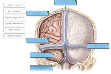
Table of Contents
Label the Cranial Dura Septa in the Figure: A Comprehensive Guide
The cranial dura mater, the outermost layer of the meninges, is not a simple, continuous sheet. Instead, it forms several important septa—folds of dura—that compartmentalize the cranial cavity, providing structural support and protection for the brain. Accurately labeling these septa is crucial for understanding brain anatomy and its clinical implications. This comprehensive guide will delve into the identification and function of each cranial dura septum, supplemented by detailed descriptions to aid in accurate labeling of any provided figure.
Understanding the Cranial Dura Mater and its Septa
Before we dive into labeling the septa, let's establish a foundational understanding of the dura mater itself. The dura mater is a tough, fibrous membrane composed of two layers: the periosteal layer (external) and the meningeal layer (internal). These layers are typically fused, except where they separate to form the dural septa. These septa are crucial for:
- Compartmentalization: Dividing the cranial cavity into distinct compartments, limiting the spread of intracranial hemorrhage or infection.
- Support: Providing structural support to the brain, preventing excessive movement.
- Protection: Acting as a protective barrier against trauma.
The Major Cranial Dura Septa: Identification and Function
Now, let's explore the major dural septa, focusing on their characteristics that aid in accurate identification within any anatomical figure:
1. Falx Cerebri: The Great Longitudinal Dividing Wall
The falx cerebri is the largest of the dural septa. Its name, literally meaning "sickle of the brain," describes its characteristic crescent shape. This large, sickle-shaped fold projects vertically downwards from the crista galli of the ethmoid bone and the internal occipital crest. It runs along the longitudinal fissure, separating the two cerebral hemispheres.
Key features for identification:
- Crescent shape: The most distinguishing feature.
- Attachment points: The crista galli anteriorly and the internal occipital crest posteriorly.
- Superior sagittal sinus: The superior sagittal sinus is located within the superior border of the falx cerebri. This sinus is a crucial component of the venous drainage system of the brain.
- Inferior sagittal sinus: A smaller sinus, located inferiorly, is also often associated with the falx cerebri.
2. Tentorium Cerebelli: The Tent Over the Cerebellum
The tentorium cerebelli is a large, tent-like structure that separates the occipital lobes of the cerebrum from the cerebellum. Its name, meaning "tent of the cerebellum," accurately reflects its function. This septum is particularly important because it protects the cerebellum from upward displacement.
Key features for identification:
- Tent-like structure: Its arched shape differentiates it from other septa.
- Attachment points: It attaches to the petrous portion of the temporal bones, the occipital bone, and the clinoid processes of the sphenoid bone.
- Transverse sinus: The transverse sinuses run within the posterior border of the tentorium. These sinuses are major venous channels draining the brain.
- Straight sinus: Located where the falx cerebri and tentorium cerebelli meet.
3. Falx Cerebelli: The Smaller Longitudinal Dividing Wall
The falx cerebelli is a smaller, sickle-shaped fold that separates the two cerebellar hemispheres. Unlike the falx cerebri, it is much smaller and less prominent. It occupies the vermis of the cerebellum.
Key features for identification:
- Smaller size: Significantly smaller than the falx cerebri.
- Location: Situated in the posterior cranial fossa, separating the cerebellar hemispheres.
- Occipital sinus: This sinus often runs within the inferior border of the falx cerebelli.
4. Diaphragma Sellae: The Roof of the Sella Turcica
The diaphragma sellae is a small, circular dural fold that forms a roof over the sella turcica, the bony depression in the sphenoid bone that houses the pituitary gland. This septum helps to protect the pituitary gland.
Key features for identification:
- Small, circular shape: Its compact size and distinct circular form are key identifiers.
- Location: Directly superior to the sella turcica.
- Opening for pituitary stalk: A small opening in the center allows for the passage of the pituitary stalk, which connects the hypothalamus to the pituitary gland.
Clinical Significance of the Dural Septa
Understanding the dural septa is crucial in various clinical settings. Their integrity plays a vital role in protecting the brain from injury and infection:
- Brain herniation: Increased intracranial pressure can force brain tissue to herniate through openings in the dural septa. For example, uncal herniation occurs when the medial temporal lobe herniates through the tentorial notch.
- Subdural hematoma: Bleeding between the dura mater and arachnoid mater can cause a subdural hematoma. The septa can influence the spread of this bleeding.
- Intracranial infections: Dural septa can limit the spread of infections within the brain, but breaches in these septa can facilitate wider dissemination.
Practical Application: Labeling a Figure
When labeling a figure depicting the cranial dura septa, carefully consider the following:
- Shape and location: Focus on the characteristic shape and location of each septum.
- Adjacent structures: Observe the relationships between the septa and surrounding brain structures.
- Sinuses: Identify the major dural sinuses associated with each septum.
- Consistent labeling: Use clear and consistent labeling to avoid confusion.
By systematically considering these aspects, you'll be able to accurately label the cranial dura septa in any anatomical illustration. Always double-check your work against anatomical atlases or textbooks to ensure accuracy.
Advanced Considerations and Related Structures
While the four major septa are the focus of most introductory anatomical studies, understanding related structures enhances comprehensive comprehension.
- Superior and Inferior Petrosal Sinuses: These sinuses, closely associated with the temporal bone and tentorium cerebelli, contribute significantly to cerebral venous drainage.
- Cavernous Sinus: This complex venous sinus, located on either side of the sella turcica, houses several cranial nerves and the internal carotid artery. Understanding its relationship to the diaphragma sellae is crucial.
- Clivus: The bony surface of the skull base to which several structures attach, influencing dural relationships.
Conclusion: Mastering the Anatomy of Cranial Dura Septa
The accurate labeling of cranial dura septa requires a thorough understanding of their anatomy, function, and clinical significance. This guide provides a comprehensive overview of these vital structures, equipping you to confidently label any anatomical figure and apply this knowledge to clinical scenarios. By mastering the identification of the falx cerebri, tentorium cerebelli, falx cerebelli, and diaphragma sellae, you will lay a solid foundation for further exploration of the intricate anatomy of the brain and its protective layers. Remember to consult reliable anatomical resources to reinforce your understanding and refine your labeling skills. Consistent practice and review are key to mastering this complex but essential aspect of neuroanatomy.
Latest Posts
Latest Posts
-
Dividing Polynomials Math Lib Answer Key
Mar 31, 2025
-
What Is Held Constant In Gay Lussacs Law
Mar 31, 2025
-
Example Of A Line In A Poem
Mar 31, 2025
-
Interval Of Convergence Of A Taylor Series
Mar 31, 2025
Related Post
Thank you for visiting our website which covers about Label The Cranial Dura Septa In The Figure. . We hope the information provided has been useful to you. Feel free to contact us if you have any questions or need further assistance. See you next time and don't miss to bookmark.
