Label The Features Of The Thoracic Cage
Muz Play
Mar 26, 2025 · 6 min read
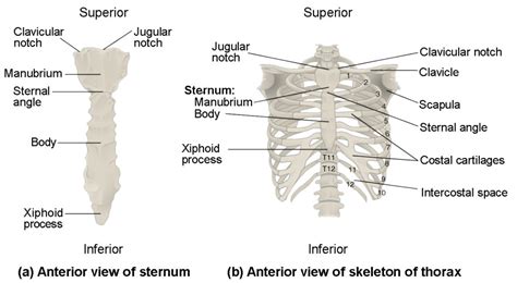
Table of Contents
Labeling the Features of the Thoracic Cage: A Comprehensive Guide
The thoracic cage, also known as the rib cage, is a bony structure of vital importance, protecting delicate organs like the heart and lungs while playing a crucial role in respiration. Understanding its intricate anatomy is fundamental for medical professionals, anatomy students, and anyone interested in human biology. This comprehensive guide will delve into the detailed labeling of the thoracic cage's features, providing a thorough understanding of its components and their functions. We'll explore the bones, joints, and associated structures, ensuring a complete and robust overview.
The Sternum: The Breastbone's Key Features
The sternum, or breastbone, is a flat bone located in the anterior midline of the thoracic cage. It's divided into three main parts:
1. Manubrium: The Handle
The manubrium sterni is the superior portion of the sternum. Its key features include:
- Jugular Notch (Suprasternal Notch): A palpable indentation at the superior border of the manubrium. It's a useful landmark for locating the second rib, crucial in clinical examinations.
- Clavicular Notches: Located on either side of the jugular notch, these articulate with the medial ends of the clavicles (collarbones), forming the sternoclavicular joints.
- Sternal Angle (Angle of Louis): A palpable ridge formed by the junction of the manubrium and the body of the sternum. This is another critical landmark, lying at the level of the second costal cartilage, useful for counting ribs during physical examinations and locating important underlying structures.
2. Body: The Main Section
The body of the sternum is the longest portion, articulating with the costal cartilages of ribs 2 through 7. Its features are less prominent than the manubrium but equally crucial:
- Costal Cartilage Articulations: These are the points of connection between the sternum and the costal cartilages, allowing for the flexibility of the rib cage during breathing. The articulation with the second rib is particularly significant due to its use as a landmark.
- Transverse Ridges: These faint ridges on the anterior surface indicate the sites of fusion of the original sternebrae (embryonic segments of the sternum).
3. Xiphoid Process: The Tip
The xiphoid process is the smallest and most inferior part of the sternum. It's cartilaginous in youth but ossifies (turns to bone) with age. Its features include:
- Cartilaginous Nature (in youth): This initial cartilaginous nature allows for flexibility during development.
- Ossification in Adulthood: The process of turning to bone, important for structural integrity in later life.
- Attachment Point: Provides attachment points for some abdominal muscles. Its location, just below the diaphragm, makes it a landmark for various procedures.
The Ribs: The Protective Framework
Twelve pairs of ribs form the lateral walls of the thoracic cage. They are classified into three groups based on their anterior attachments:
1. True Ribs (Ribs 1-7): Direct Connection
These ribs articulate directly with the sternum via their own costal cartilages. The features of a typical true rib include:
- Head: Articulates with the vertebral bodies of two adjacent thoracic vertebrae.
- Neck: The constricted portion connecting the head and tubercle.
- Tubercle: A small projection that articulates with the transverse process of a vertebra.
- Angle: A sharp bend in the rib's shaft.
- Shaft (Body): The long, curved portion of the rib.
- Costal Groove: A groove on the inferior border, housing the intercostal vessels and nerves.
2. False Ribs (Ribs 8-10): Indirect Connection
These ribs don't directly articulate with the sternum; instead, their costal cartilages fuse to the costal cartilage of the rib above, forming a chain connection to the sternum. They share the same features as true ribs.
3. Floating Ribs (Ribs 11-12): No Anterior Connection
These ribs are the shortest and lack anterior attachments to the sternum or other costal cartilages. Their features also mirror those of true ribs, however, their lack of anterior connection makes them more mobile.
The Vertebrae: The Posterior Support
The thoracic vertebrae (T1-T12) form the posterior aspect of the thoracic cage. They differ slightly from cervical and lumbar vertebrae:
- Heart-Shaped Body: Larger than cervical vertebrae, providing increased stability.
- Costal Facets: Articulation sites on the vertebral bodies and transverse processes for rib articulation. These facets are crucial for the rib cage's structure.
- Spinous Processes: Long and pointed, projecting downwards, which are easily palpable.
- Vertebral Foramina: Openings for the spinal cord.
- Intervertebral Foramina: Openings between adjacent vertebrae for spinal nerves.
Each thoracic vertebra articulates with ribs through the costal facets, contributing to the intricate articulation and movement of the ribcage.
Joints of the Thoracic Cage: Articulations for Movement and Stability
Several types of joints contribute to the thoracic cage's mobility and stability:
- Costovertebral Joints: Articulations between the head of a rib and the bodies of two adjacent thoracic vertebrae.
- Costotransverse Joints: Articulations between the tubercle of a rib and the transverse process of a vertebra.
- Sternoclavicular Joints: Articulations between the clavicles and the manubrium of the sternum.
- Costochondral Joints: Articulations between the ribs and their respective costal cartilages.
- Interchondral Joints: Articulations between adjacent costal cartilages.
- Sternocostal Joints: Articulations between the costal cartilages and the sternum.
These joints, together with ligaments and muscles, allow for the expansion and contraction of the chest cavity during breathing.
Associated Structures: Muscles and Vessels
The thoracic cage isn't just a bony structure; it's intertwined with muscles and vasculature crucial for its function and the body's overall health:
Muscles:
- Intercostal Muscles: Located between the ribs, these muscles play a vital role in respiration, assisting with inhalation and exhalation.
- Diaphragm: A dome-shaped muscle separating the thoracic and abdominal cavities, it's the primary muscle of respiration.
- Other Muscles: Numerous other muscles attach to the thoracic cage, including those involved in shoulder movement, posture, and trunk stability.
Vessels:
- Intercostal Arteries and Veins: These blood vessels run along the intercostal spaces, supplying blood to the thoracic wall.
- Internal Thoracic Arteries: These blood vessels run down the anterior thoracic wall, supplying blood to various structures.
The precise interaction between these muscles and blood vessels is essential for the normal functioning of the respiratory system.
Clinical Significance: Why Understanding the Thoracic Cage Matters
A thorough understanding of the thoracic cage is vital in numerous medical fields:
- Cardiology: The thoracic cage protects the heart; knowing its anatomy is fundamental for cardiac procedures and diagnoses.
- Pulmonology: Lung function is intrinsically linked to the rib cage's movement; understanding its anatomy is crucial for diagnosing and treating respiratory conditions.
- Trauma Surgery: Rib fractures and other injuries to the thoracic cage are common, requiring a deep understanding of its anatomy for effective treatment.
- Orthopedics: Conditions affecting the joints and bones of the thoracic cage require specialized knowledge for diagnosis and management.
Conclusion: A Detailed Roadmap to the Thoracic Cage
This detailed guide has provided a comprehensive overview of the thoracic cage's features. From the sternum's components and the varied types of ribs to the intricate joints and associated structures, understanding each element is vital for a complete grasp of human anatomy and its clinical implications. The information provided here serves as a robust foundation for further exploration and study, emphasizing the critical role of this protective and dynamic structure in human health. Remembering the key landmarks, such as the sternal angle and jugular notch, facilitates accurate anatomical location and clinical assessment. Continued learning and reference to anatomical resources will reinforce this knowledge and enhance its practical application.
Latest Posts
Latest Posts
-
Particles That Are Too Big For Diffusion And Active Transport
Mar 29, 2025
-
Phonology Morphology Syntax Semantics And Pragmatics
Mar 29, 2025
-
Turbulent Channel Flow Near Wall Velocity Profile
Mar 29, 2025
-
Essentials Of Oceanography Pdf Free Download
Mar 29, 2025
-
Lyrics Of Ode To Billy Joe
Mar 29, 2025
Related Post
Thank you for visiting our website which covers about Label The Features Of The Thoracic Cage . We hope the information provided has been useful to you. Feel free to contact us if you have any questions or need further assistance. See you next time and don't miss to bookmark.
