Label The Indicated Muscles Of The Head And Neck
Muz Play
Mar 24, 2025 · 6 min read
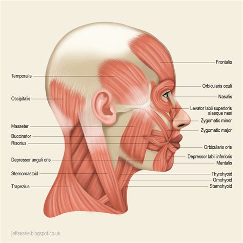
Table of Contents
Label the Indicated Muscles of the Head and Neck: A Comprehensive Guide
Understanding the intricate musculature of the head and neck is crucial for anyone studying anatomy, physiotherapy, or related fields. This region boasts a complex network of muscles responsible for facial expression, chewing, swallowing, head movement, and more. This detailed guide will walk you through identifying and understanding the key muscles of the head and neck, providing a comprehensive overview perfect for students and professionals alike. We'll explore their origins, insertions, actions, and clinical relevance.
Major Muscles of the Head and Neck: A Categorized Approach
For clarity, we'll categorize the muscles based on their location and function. Remember, accurate labeling requires a strong grasp of anatomical terminology and spatial relationships. Visual aids like anatomical charts and models are highly recommended throughout this learning process.
Muscles of Facial Expression: Shaping Emotion and Communication
The muscles of facial expression are unique in that they are directly attached to the skin, allowing for a wide range of movements that convey our emotions and facilitate communication.
1. Frontalis: Located on the forehead, this muscle raises the eyebrows, creating wrinkles on the forehead (think surprise or concern). Origin: Galea aponeurotica. Insertion: Skin of the eyebrows. Action: Elevates eyebrows, wrinkles forehead.
2. Orbicularis Oculi: This circular muscle surrounds the eye. It's responsible for closing the eyelids, squinting, and protecting the eye from foreign objects. Origin: Medial palpebral ligament and surrounding bones. Insertion: Skin around the eyelids. Action: Closes eyelids, squints.
3. Orbicularis Oris: This muscle forms the lips. Its complex structure allows for a variety of movements, including pursing, kissing, and whistling. Origin: Maxilla and mandible. Insertion: Skin and mucous membrane of the lips. Action: Compresses and protrudes lips.
4. Zygomaticus Major and Minor: These muscles originate from the zygomatic bone (cheekbone) and insert into the corner of the mouth. They are responsible for smiling and elevating the corners of the mouth. Origin: Zygomatic bone. Insertion: Angle of the mouth. Action: Elevates the corners of the mouth (smiling).
5. Buccinator: This flat muscle forms the cheek. It plays a role in chewing, whistling, and sucking. Origin: Maxilla and mandible. Insertion: Orbicularis oris. Action: Compresses cheeks, assists in mastication.
6. Masseter: While technically a muscle of mastication, the masseter is visible on the side of the face and contributes to facial contours. It is a powerful muscle responsible for closing the jaw. Origin: Zygomatic arch. Insertion: Angle of the mandible. Action: Elevates mandible (closes jaw).
7. Temporalis: Another muscle of mastication, the temporalis lies on the side of the head, fanning out towards the temporal fossa. It also plays a significant role in closing the jaw. Origin: Temporal fossa. Insertion: Coronoid process of the mandible. Action: Elevates and retracts mandible.
Muscles of Mastication: The Power Behind Chewing
These muscles are responsible for the complex movements involved in chewing (mastication).
1. Masseter (already described above)
2. Temporalis (already described above)
3. Medial Pterygoid: This deep muscle lies inside the mouth, close to the pterygoid plates. It assists in closing and protracting the jaw. Origin: Medial surface of the lateral pterygoid plate. Insertion: Medial surface of the angle of the mandible. Action: Elevates and protracts mandible.
4. Lateral Pterygoid: This muscle also contributes to jaw movement, playing a key role in opening and lateral movement (side-to-side chewing). Origin: Greater wing and lateral pterygoid plate of the sphenoid bone. Insertion: Neck of the mandible and articular disc of the temporomandibular joint. Action: Depresses and protracts mandible, lateral movement.
Muscles of the Neck: Supporting Head Movement and Posture
The muscles of the neck support the weight of the head, allow for a wide range of movement, and contribute to overall posture.
1. Sternocleidomastoid (SCM): A prominent muscle that extends from the sternum and clavicle to the mastoid process of the temporal bone. It is crucial for head rotation and flexion. Origin: Manubrium of the sternum and medial portion of the clavicle. Insertion: Mastoid process of the temporal bone. Action: Flexes neck, rotates head.
2. Trapezius: A large, superficial muscle that covers much of the upper back and neck. It plays a vital role in shoulder movement and head extension. Origin: Occipital bone, nuchal ligament, and spinous processes of C7-T12 vertebrae. Insertion: Lateral third of the clavicle, acromion, and spine of the scapula. Action: Elevates, depresses, retracts, and rotates scapula; extends head.
3. Scalenes (Anterior, Middle, Posterior): These three muscles are located deep in the neck, flanking the vertebral column. They play a crucial role in neck flexion and respiration. Origin: Transverse processes of cervical vertebrae. Insertion: Ribs 1 and 2. Action: Flexes neck, elevates ribs (assists in respiration).
4. Splenius Capitis and Cervicis: These paired muscles are deep to the trapezius, extending from the cervical and thoracic vertebrae to the occipital bone and cervical vertebrae. They are involved in head extension and rotation. Origin: Spinous processes of thoracic and cervical vertebrae. Insertion: Occipital bone and transverse processes of upper cervical vertebrae. Action: Extends and rotates head.
5. Longus Capitis and Colli: These deep neck flexor muscles run along the anterior aspect of the vertebral column, contributing to head and neck flexion. Origin: Anterior surfaces of cervical vertebrae. Insertion: Anterior aspect of the occipital bone and cervical vertebrae. Action: Flexes head and neck.
Clinical Relevance: Understanding the Implications
Knowledge of the head and neck muscles is critical in various clinical settings. For example:
-
Temporomandibular Joint (TMJ) Disorders: Understanding the muscles of mastication is vital for diagnosing and treating TMJ disorders, which involve pain and dysfunction of the jaw joint.
-
Cervical Spine Injuries: Knowledge of neck musculature is crucial in assessing and managing injuries to the cervical spine, which can range from whiplash to more severe fractures.
-
Facial Nerve Palsy (Bell's Palsy): Understanding the muscles of facial expression is essential for diagnosing and managing Bell's palsy, a condition causing facial weakness or paralysis.
-
Headaches and Migraines: Tension headaches and migraines can be linked to muscle tension in the neck and head, highlighting the importance of understanding these muscles in treating these conditions.
-
Swallowing Disorders (Dysphagia): Muscles involved in swallowing (including some of the muscles of the head and neck) need to be assessed to understand and treat dysphagia, which is difficulty swallowing.
Practical Tips for Effective Muscle Identification:
-
Utilize Anatomical Models and Charts: Visual aids are indispensable for understanding the complex relationships between muscles.
-
Palpate Muscles: Gently touch and feel the muscles during various movements to better understand their location and action. (Always obtain permission before touching another person.)
-
Practice Regularly: Consistent practice is key to mastering the identification of these muscles. Review the information frequently, utilizing flashcards or other memory aids.
-
Relate Structure to Function: Understanding how a muscle's structure influences its action is essential for truly grasping the material.
-
Learn in Layers: Begin by identifying the superficial muscles and then progressively work your way to the deeper structures.
Conclusion: A Foundation for Deeper Understanding
This comprehensive guide has provided an overview of the muscles of the head and neck. By understanding their origins, insertions, actions, and clinical significance, you'll have built a strong foundation for further study in anatomy and related fields. Remember, continuous practice and the utilization of visual resources are key to successfully mastering the identification of these crucial muscles. Continue your studies with detailed anatomical atlases and consider exploring more advanced resources as your understanding grows. The detailed study of head and neck muscles will reward you with a deeper appreciation for the intricate workings of the human body.
Latest Posts
Latest Posts
-
What Is The Reactants Of Glycolysis
Mar 26, 2025
-
An Introduction To General Organic And Biological Chemistry
Mar 26, 2025
-
The Cutaneous Membrane Is Also Known As
Mar 26, 2025
-
Solve The Following System Of Equations Algebraically
Mar 26, 2025
-
Why Are Tertiary Carbocations More Stable
Mar 26, 2025
Related Post
Thank you for visiting our website which covers about Label The Indicated Muscles Of The Head And Neck . We hope the information provided has been useful to you. Feel free to contact us if you have any questions or need further assistance. See you next time and don't miss to bookmark.
