Label The Microscopic Anatomy Of Cardiac Muscle
Muz Play
Mar 21, 2025 · 6 min read
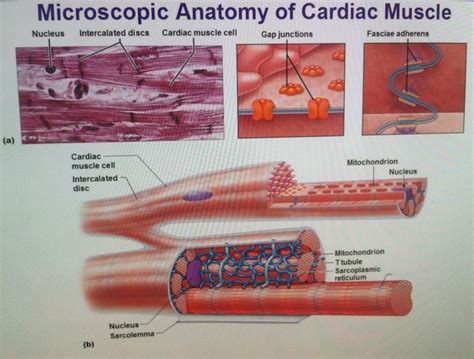
Table of Contents
Labeling the Microscopic Anatomy of Cardiac Muscle: A Comprehensive Guide
Cardiac muscle, the powerhouse of the heart, is a specialized type of muscle tissue responsible for the rhythmic contractions that pump blood throughout the body. Understanding its microscopic anatomy is crucial for comprehending the physiological processes of the heart and diagnosing cardiovascular diseases. This detailed guide will walk you through the key components of cardiac muscle at the microscopic level, providing a comprehensive labeling exercise and highlighting important functional aspects.
Key Structural Features of Cardiac Muscle Cells (Cardiomyocytes)
Cardiac muscle, unlike skeletal muscle, is composed of branched, interconnected cells called cardiomyocytes. These cells are shorter and wider than skeletal muscle fibers and are characterized by several unique features visible under a microscope:
1. Intercalated Discs: The Connecting Links
Intercalated discs are the most striking feature of cardiac muscle tissue. These are complex junctions that connect adjacent cardiomyocytes, forming a functional syncytium. Under the microscope, they appear as dark, transverse lines crossing the muscle fibers. Intercalated discs are crucial for efficient electrical coupling and mechanical adhesion between cells. They consist of several specialized components:
- Fascia adherens: These act as anchoring sites for actin filaments, transmitting the force of contraction from one cell to the next. Think of them as the "glue" holding the cells together mechanically.
- Maculae adherentes (desmosomes): Providing strong adhesion between cells, desmosomes prevent the cells from separating during contraction. They act like rivets, reinforcing the structure under stress.
- Gap junctions: These are communication channels that allow the rapid spread of electrical impulses between adjacent cardiomyocytes. This ensures coordinated and synchronous contractions of the heart muscle. They are vital for the heart's ability to function as a single, coordinated unit.
2. Sarcomeres: The Contractile Units
Like skeletal muscle, cardiac muscle is organized into sarcomeres, the basic contractile units. These are repeating units of overlapping actin and myosin filaments arranged in a highly organized pattern. Under the microscope, the striated appearance of cardiac muscle is due to the regular arrangement of these sarcomeres. The banding pattern consists of:
- Z-lines: These define the boundaries of each sarcomere. They appear as dark lines under the microscope.
- I-bands: These are light bands containing only thin actin filaments.
- A-bands: These are dark bands containing both thick myosin and thin actin filaments.
- H-zone: This is a lighter region within the A-band where only thick myosin filaments are present.
- M-line: This is a dark line in the center of the H-zone, anchoring myosin filaments.
3. Myofibrils: Bundles of Sarcomeres
Multiple sarcomeres are arranged end-to-end to form myofibrils, long cylindrical structures running the length of the cardiomyocyte. These myofibrils are responsible for the muscle cell's contractile properties. The arrangement of myofibrils within the cell contributes to the overall striated appearance of the tissue.
4. Mitochondria: Powerhouses of the Heart
Cardiac muscle cells are highly energy-demanding, requiring a constant supply of ATP for contraction. This is reflected in the abundance of mitochondria, the cell's energy-producing organelles, within cardiomyocytes. Under the microscope, mitochondria appear as numerous, elongated structures scattered throughout the cytoplasm. Their high number underlines the heart's continuous need for energy.
5. T-Tubules (Transverse Tubules): Inward Extensions of the Sarcolemma
T-tubules are invaginations of the sarcolemma (cell membrane) that extend deep into the muscle cell, reaching the sarcomeres. They are involved in the rapid transmission of electrical signals from the sarcolemma to the sarcoplasmic reticulum, triggering muscle contraction. They are less extensive than in skeletal muscle.
6. Sarcoplasmic Reticulum (SR): Calcium Storage
The sarcoplasmic reticulum (SR) is a specialized network of intracellular membranes that store and release calcium ions (Ca²⁺). Ca²⁺ is essential for muscle contraction, and the SR plays a critical role in regulating its availability. The SR in cardiac muscle is less developed than in skeletal muscle.
7. Nuclei: The Control Centers
Cardiac muscle cells are typically uninucleated, although some may contain two nuclei. The nuclei are centrally located and are easily identifiable under the microscope.
Labeling Exercise: A Step-by-Step Guide
To solidify your understanding, let's go through a labeling exercise. Imagine you are observing a microscopic image of cardiac muscle tissue. You should be able to identify and label the following structures:
- Cardiomyocyte: The individual muscle cell.
- Intercalated Disc: The dark, transverse line connecting adjacent cardiomyocytes. Label its components:
- Fascia adherens: The anchoring site for actin filaments.
- Desmosomes (Maculae adherentes): Providing strong adhesion between cells.
- Gap junctions: Channels for electrical impulse transmission.
- Sarcomere: The basic contractile unit. Label its components:
- Z-line: Boundary of the sarcomere.
- I-band: Light band with only actin filaments.
- A-band: Dark band with both actin and myosin filaments.
- H-zone: Lighter region within the A-band with only myosin filaments.
- M-line: Center of the H-zone, anchoring myosin filaments.
- Myofibrils: Long cylindrical structures containing sarcomeres.
- Mitochondria: Numerous, elongated organelles responsible for energy production.
- T-tubules: Invaginations of the sarcolemma.
- Sarcoplasmic Reticulum (SR): Network of membranes storing and releasing calcium ions.
- Nucleus: The cell's control center (usually centrally located).
Clinical Significance: Understanding the Implications
Understanding the microscopic anatomy of cardiac muscle is paramount in various clinical contexts:
- Cardiomyopathies: Diseases affecting the heart muscle, such as hypertrophic cardiomyopathy or dilated cardiomyopathy, often involve structural changes within cardiomyocytes and their connections (intercalated discs). Microscopic examination can reveal these abnormalities.
- Myocardial Infarction (Heart Attack): Microscopic analysis of heart tissue post-heart attack reveals the extent of muscle cell damage and necrosis, crucial for determining the severity of the injury and prognosis.
- Cardiac Arrhythmias: Disruptions in the electrical conduction system of the heart, reflected in the function of gap junctions within intercalated discs, can lead to various arrhythmias. Microscopic studies may not directly show these issues but aid in understanding the underlying cellular pathology.
- Drug Development: Research into new drugs for cardiovascular diseases often involves studying their effects on the microscopic structure and function of cardiac muscle cells.
Conclusion: A Microscopic View of a Vital Organ
The microscopic anatomy of cardiac muscle is far more complex than a simple glance might suggest. The intricate arrangement of cardiomyocytes, their specialized junctions, and the abundance of energy-producing organelles underscore the heart's relentless work. By understanding these details, we gain a deeper appreciation for the heart's remarkable physiology and the implications for cardiovascular health and disease. The labeling exercise provided above should serve as a valuable tool for reinforcing your understanding of this complex and fascinating tissue. Further study and exploration through microscopy are highly encouraged for a complete comprehension.
Latest Posts
Latest Posts
-
The Basic Unit Of Life Is
Mar 21, 2025
-
When An Atom Gains An Electron It Becomes
Mar 21, 2025
-
What Percentage Of Urine Is Water
Mar 21, 2025
-
What Are Some Nutrients That Plants Need
Mar 21, 2025
-
The Monomer Of A Nucleic Acid
Mar 21, 2025
Related Post
Thank you for visiting our website which covers about Label The Microscopic Anatomy Of Cardiac Muscle . We hope the information provided has been useful to you. Feel free to contact us if you have any questions or need further assistance. See you next time and don't miss to bookmark.
