Label The Parts Of A Gomphosis
Muz Play
Mar 24, 2025 · 6 min read
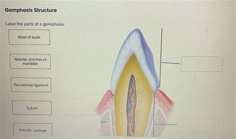
Table of Contents
Delving Deep: A Comprehensive Guide to Labeling the Parts of a Gomphosis
Gomphoses, a fascinating type of fibrous joint, are often overlooked in discussions of the skeletal system. However, understanding their unique structure and function is crucial for comprehending the overall mechanics of the body, particularly concerning the cranium and dentition. This comprehensive guide will provide a detailed exploration of gomphoses, focusing on the identification and labeling of their key components. We'll explore their defining characteristics, their role in supporting the teeth, and the subtle yet significant differences compared to other types of fibrous joints.
What is a Gomphosis?
A gomphosis is a fibrous joint characterized by the peg-in-socket articulation. This unique type of joint is found only where teeth articulate with their sockets in the alveolar processes of the maxilla and mandible. The word "gomphosis" itself originates from the Greek word "gomphos," meaning "bolt" or "peg," aptly describing the structure's resemblance to a nail or peg firmly fixed in place. Unlike other fibrous joints like sutures or syndesmoses, which connect bones to bones, a gomphosis connects a tooth (a specialized structure, not technically a bone) to the bone of the jaw.
The key characteristic differentiating a gomphosis from other joint types is the specialized connective tissue known as the periodontal ligament. This ligament is responsible for anchoring the tooth within its socket, providing stability, and enabling slight movement crucial for shock absorption and sensory feedback during mastication (chewing).
Labeling the Parts of a Gomphosis: A Detailed Breakdown
Let's break down the components of a gomphosis and understand their individual roles:
1. The Tooth (Dens):
- This is the "peg" in the peg-in-socket articulation. The tooth is comprised of several distinct parts:
- Enamel: The hard, outermost layer of the crown, providing protection.
- Dentin: The layer underlying the enamel, forming the bulk of the tooth structure.
- Cementum: A bone-like substance covering the root of the tooth, anchoring the periodontal ligament.
- Pulp Cavity: The central core of the tooth containing blood vessels, nerves, and connective tissue, providing nourishment and sensation.
- Root: The portion of the tooth embedded within the alveolar socket. The root's surface is crucial for the attachment of the periodontal ligament.
2. The Alveolar Socket (Alveolus):
- This is the "socket" within which the tooth is embedded. It is a bony cavity within the maxilla (upper jaw) or mandible (lower jaw). The alveolar bone is highly specialized, adapted to support the forces generated during chewing.
3. The Periodontal Ligament (PDL):
- This is the critical component defining the gomphosis as a distinct joint type. The PDL is a dense connective tissue composed of collagen fibers, fibroblasts, osteoblasts, osteoclasts, and cementoblasts. It acts as a crucial interface between the tooth and the alveolar bone, performing several essential functions:
- Attachment: Collagen fibers within the PDL firmly attach the cementum of the root to the alveolar bone, anchoring the tooth securely. These fibers are arranged in distinct bundles, creating a resilient network.
- Shock Absorption: The PDL acts as a shock absorber, distributing forces generated during mastication and preventing damage to the tooth and alveolar bone.
- Sensory Perception: The PDL contains sensory nerve endings, allowing for the perception of pressure, pain, and proprioception (awareness of tooth position).
- Nutrient Supply: The PDL facilitates the delivery of nutrients and oxygen to the tooth and the removal of metabolic waste products.
- Remodeling: The PDL plays a role in the continuous remodeling and adaptation of the alveolar bone and cementum in response to functional demands. This ensures the stability of the tooth throughout life.
4. Cementum:
- This hard, bone-like tissue covers the root of the tooth. It is crucial for the attachment of the periodontal ligament fibers, providing a strong and resilient interface with the alveolar bone. Cementum's role is critical in the overall stability of the tooth within its socket.
5. Alveolar Bone (Alveolar Process):
- This is the specialized bone forming the socket. It's highly vascularized and innervated, reflecting its critical role in maintaining the health and integrity of the gomphosis. The alveolar bone is constantly remodeled in response to forces exerted on the teeth.
Comparing Gomphosis to Other Fibrous Joints
While gomphoses are classified as fibrous joints, it's essential to distinguish them from other types:
- Sutures: Sutures are fibrous joints connecting the bones of the skull. They are characterized by interlocking edges, with very little movement.
- Syndesmoses: Syndesmoses are fibrous joints where bones are connected by a ligament or a sheet of fibrous tissue, allowing for a small degree of movement. Examples include the distal tibiofibular joint.
The key difference lies in the function and location: gomphoses are specialized for the unique demands of tooth implantation and function, unlike sutures and syndesmoses which connect bones solely. The presence of the periodontal ligament as the primary connecting tissue is the definitive characteristic distinguishing gomphosis from other fibrous joints.
Clinical Significance of Understanding Gomphosis
Understanding the structure and function of the gomphosis is crucial in several clinical contexts:
- Periodontal Disease: Periodontal disease, a common inflammatory condition, affects the periodontal ligament and alveolar bone. This can lead to tooth loosening and eventual loss. Understanding the anatomy is key to diagnosis and treatment.
- Orthodontics: Orthodontic treatments aim to reposition teeth. This requires a thorough understanding of the forces exerted on the periodontal ligament and alveolar bone to achieve safe and effective tooth movement.
- Implantology: Dental implants rely on the principle of osseointegration, where the implant integrates with the alveolar bone, mimicking the natural stability of a gomphosis. Knowledge of gomphosis anatomy is vital for successful implant placement.
- Trauma: Injuries to the teeth and jaws can damage the gomphosis, leading to tooth mobility or avulsion (complete displacement). Understanding the structure helps in diagnosis and treatment planning.
Advanced Considerations: Microscopic Anatomy and Biomechanics
Delving deeper, the microscopic anatomy of the periodontal ligament reveals a complex arrangement of collagen fibers. These fibers are not uniformly oriented but rather organized into distinct bundles, including:
- Oblique Fibers: The most numerous, running obliquely from the cementum to the alveolar bone. They resist occlusal forces (forces generated during chewing).
- Transseptal Fibers: Connecting adjacent teeth, enhancing the interdental support.
- Apical Fibers: Running from the apex (tip) of the root to the alveolar bone.
- Horizontal Fibers: Running horizontally between the cementum and alveolar bone.
- Interradicular Fibers: Found between the roots of multi-rooted teeth.
The biomechanics of the gomphosis are equally complex, involving the interaction of various tissues under the influence of occlusal forces. The periodontal ligament's ability to adapt to these forces over time contributes to the remarkable stability and longevity of the tooth in its socket. Research into these areas continues to refine our understanding of this critical joint type.
Conclusion: The Gomphosis – A Vital Yet Often Underappreciated Joint
The gomphosis, a specialized fibrous joint connecting teeth to the alveolar bone, showcases a remarkable adaptation for mastication and sensory feedback. Understanding its components – the tooth, alveolar socket, periodontal ligament, cementum, and alveolar bone – is crucial for comprehending the intricate mechanics of the craniofacial region and for addressing various clinical scenarios involving the teeth and surrounding structures. The detailed exploration of its microscopic anatomy and biomechanics provides a deeper appreciation of the intricate design and functional efficiency of this often-overlooked yet vital joint. The continued research in this area constantly reveals new insights into the complex interplay between tissues, making the study of the gomphosis an ongoing and fascinating endeavor. This deeper understanding not only enhances our comprehension of basic biology but also greatly benefits the field of dentistry and oral health.
Latest Posts
Latest Posts
-
Newspapers During The Revolutionary War Period Tended To
Mar 25, 2025
-
Are Ketones Or Aldehydes More Reactive
Mar 25, 2025
-
A Unicellular Protist Is Part Of Which Domain
Mar 25, 2025
-
A Species With 12 Protons And 10 Electrons Is
Mar 25, 2025
-
Which Organelles Are Found Only In Plant Cells
Mar 25, 2025
Related Post
Thank you for visiting our website which covers about Label The Parts Of A Gomphosis . We hope the information provided has been useful to you. Feel free to contact us if you have any questions or need further assistance. See you next time and don't miss to bookmark.
