Microscopic Anatomy And Organization Of Skeletal Muscle
Muz Play
Apr 02, 2025 · 7 min read

Table of Contents
Microscopic Anatomy and Organization of Skeletal Muscle: A Deep Dive
Skeletal muscle, the powerhouse of voluntary movement, is a fascinating marvel of biological engineering. Understanding its microscopic anatomy and intricate organization is key to comprehending how we move, maintain posture, and generate force. This detailed exploration will delve into the structural hierarchy of skeletal muscle, from the macroscopic to the molecular level, examining the key components that contribute to its remarkable functionality.
The Hierarchical Structure of Skeletal Muscle
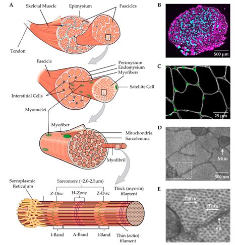
Skeletal muscle's organization is hierarchical, with each level contributing specific properties to the overall function. This organized structure allows for efficient force generation and transmission.
1. Muscle Fiber (Muscle Cell): The Fundamental Unit
The fundamental unit of skeletal muscle is the muscle fiber, also known as a myofiber. These are long, cylindrical cells, multinucleated and incredibly specialized for contraction. Their diameter can vary, ranging from 10 to 100 micrometers, and their length can be astonishing, sometimes reaching the entire length of the muscle itself. This length is achieved through the fusion of numerous myoblasts during development.
Key Features of Muscle Fibers:
- Multinucleated: Unlike most cells, muscle fibers possess multiple nuclei located just beneath the sarcolemma (cell membrane). This reflects their origin from the fusion of multiple myoblasts.
- Sarcolemma: The plasma membrane of the muscle fiber. It plays a crucial role in transmitting the nerve impulse for muscle contraction.
- Sarcoplasm: The cytoplasm of the muscle fiber. It contains the myofibrils, the organelles responsible for contraction, as well as glycogen granules (energy storage) and myoglobin (oxygen storage).
- Myofibrils: These are the cylindrical structures running the length of the muscle fiber, representing the contractile elements. They are highly organized arrays of actin and myosin filaments, arranged in repeating units called sarcomeres.
2. Myofibrils: The Contractile Machines
Myofibrils are the powerhouses of muscle contraction. They are made up of repeating units called sarcomeres, the basic functional units of muscle contraction. The highly organized arrangement of actin and myosin filaments within the sarcomere is responsible for the striated appearance of skeletal muscle under a microscope.
Components of the Sarcomere:
- Z-lines (Z-discs): These are protein structures that mark the boundaries of each sarcomere. Actin filaments are anchored to the Z-lines.
- A-band (Anisotropic band): This dark band represents the entire length of the myosin filaments, including the regions where actin and myosin overlap.
- I-band (Isotropic band): This light band contains only actin filaments and extends from the A-band of one sarcomere to the A-band of the adjacent sarcomere. The I-band shortens during muscle contraction.
- H-zone: This lighter region in the center of the A-band contains only myosin filaments and disappears during maximal muscle contraction.
- M-line: This is a protein structure located in the center of the H-zone, helping to hold the myosin filaments in place.
3. Actin and Myosin Filaments: The Molecular Players
The sarcomere's striated appearance is due to the precise arrangement of actin and myosin filaments. These proteins are the molecular motors responsible for generating the force of muscle contraction.
Actin Filaments: Thin filaments composed primarily of the protein actin. Troponin and tropomyosin are also associated with actin filaments and play critical roles in regulating muscle contraction.
Myosin Filaments: Thick filaments composed primarily of the protein myosin. Myosin has a head region that interacts with actin filaments, forming cross-bridges during contraction. The myosin heads possess ATPase activity, hydrolyzing ATP to provide the energy for muscle contraction.
4. Sarcoplasmic Reticulum (SR): Calcium Storage and Release
The sarcoplasmic reticulum (SR) is a specialized network of endoplasmic reticulum surrounding each myofibril. Its primary function is to store and release calcium ions (Ca²⁺), essential for initiating muscle contraction. The SR's intricate structure ensures rapid and efficient calcium delivery to the myofibrils upon stimulation.
Triads: The SR forms specialized structures called triads at the junctions of A-bands and I-bands. These triads consist of two terminal cisternae of the SR flanking a T-tubule. This arrangement facilitates rapid signal transmission from the sarcolemma to the myofibrils.
5. Transverse Tubules (T-tubules): Signal Transmission
Transverse tubules (T-tubules) are invaginations of the sarcolemma that penetrate deep into the muscle fiber, forming a network that allows rapid transmission of the nerve impulse throughout the entire muscle fiber. They are closely associated with the SR, facilitating the release of calcium ions from the SR upon stimulation.
The Neuromuscular Junction: Where Nerve Meets Muscle
Muscle contraction is initiated by a signal from the nervous system. This signal is transmitted at the neuromuscular junction (NMJ), the specialized synapse between a motor neuron and a muscle fiber.
Key Components of the NMJ:
- Motor Neuron: The nerve cell that innervates the muscle fiber.
- Synaptic Terminal (Axon Terminal): The end of the motor neuron where neurotransmitters are released.
- Synaptic Cleft: The space between the motor neuron and the muscle fiber.
- Motor End Plate: A specialized region of the sarcolemma on the muscle fiber, containing receptors for the neurotransmitter acetylcholine (ACh).
Mechanism of Neuromuscular Transmission:
- An action potential (nerve impulse) arrives at the synaptic terminal.
- This triggers the release of acetylcholine (ACh) into the synaptic cleft.
- ACh binds to receptors on the motor end plate, causing depolarization of the sarcolemma.
- This depolarization initiates an action potential that travels along the sarcolemma and into the T-tubules.
- The action potential triggers the release of calcium ions (Ca²⁺) from the sarcoplasmic reticulum.
- Ca²⁺ binds to troponin, initiating the sliding filament mechanism of muscle contraction.
The Sliding Filament Mechanism: How Muscles Contract
The sliding filament mechanism describes the process by which muscle fibers shorten. It involves the interaction between actin and myosin filaments within the sarcomere.
Steps in the Sliding Filament Mechanism:
- Cross-bridge Formation: Myosin heads bind to actin filaments, forming cross-bridges.
- Power Stroke: The myosin heads pivot, pulling the actin filaments toward the center of the sarcomere. This requires ATP hydrolysis.
- Cross-bridge Detachment: ATP binds to the myosin head, causing it to detach from the actin filament.
- Cross-bridge Reactivation: The myosin head returns to its original conformation, ready to bind to another actin filament.
This cycle of cross-bridge formation, power stroke, detachment, and reactivation continues as long as calcium ions are present and ATP is available. The coordinated action of numerous myosin heads along the length of the sarcomere results in the shortening of the muscle fiber.
Muscle Fiber Types: Variations in Contractile Properties
Skeletal muscle fibers are not all created equal. They are classified into different types based on their contractile properties, primarily their speed of contraction and their resistance to fatigue.
Three main types of muscle fibers:
- Type I (Slow-twitch, oxidative): These fibers contract slowly, have high resistance to fatigue, and are rich in mitochondria (for aerobic respiration). They are ideal for endurance activities.
- Type IIa (Fast-twitch, oxidative-glycolytic): These fibers contract faster than Type I fibers, have moderate resistance to fatigue, and utilize both aerobic and anaerobic respiration. They are well-suited for activities requiring both speed and endurance.
- Type IIb (Fast-twitch, glycolytic): These fibers contract very rapidly, have low resistance to fatigue, and rely primarily on anaerobic respiration. They are best suited for short bursts of intense activity.
Muscle Organization at the Macroscopic Level: From Fibers to Muscle
The microscopic components of skeletal muscle are organized into larger functional units at the macroscopic level. Muscle fibers are bundled together into fascicles, which are further grouped together to form the entire muscle. The arrangement of fascicles influences the muscle's overall shape and function.
Different fascicle arrangements:
- Parallel: Fibers run parallel to the long axis of the muscle (e.g., sartorius).
- Pennate: Fibers attach obliquely to a central tendon (e.g., rectus femoris).
- Convergent: Fibers converge from a broad origin to a narrow insertion (e.g., pectoralis major).
- Circular: Fibers arranged in concentric rings (e.g., orbicularis oris).
Conclusion
The microscopic anatomy and organization of skeletal muscle are remarkably complex and exquisitely adapted for their function. The intricate interplay between the sarcomere's molecular components, the sarcoplasmic reticulum's calcium handling, and the neuromuscular junction's signaling mechanism allows for precise and powerful voluntary movement. Understanding these details is vital for appreciating the marvels of human physiology and the complexities of musculoskeletal function. Further research continues to uncover the intricacies of muscle biology, with implications for understanding and treating muscle diseases and injuries, as well as improving athletic performance and rehabilitation strategies.
Latest Posts
Latest Posts
-
Shortage On Supply And Demand Graph
Apr 03, 2025
-
Place The Products And Reactants Of The Citric Acid Cycle
Apr 03, 2025
-
Red Blood Cells In Hypertonic Solution
Apr 03, 2025
-
The Echelon Form Of A Matrix Is Unique
Apr 03, 2025
-
How To Write An Equation For A Vertical Line
Apr 03, 2025
Related Post
Thank you for visiting our website which covers about Microscopic Anatomy And Organization Of Skeletal Muscle . We hope the information provided has been useful to you. Feel free to contact us if you have any questions or need further assistance. See you next time and don't miss to bookmark.
