Muscles Of The Trunk Anterior View
Muz Play
Mar 15, 2025 · 7 min read
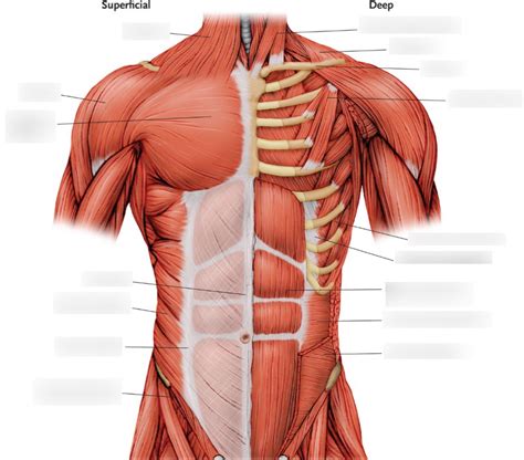
Table of Contents
Muscles of the Trunk: An Anterior View
The human trunk, the central part of the body connecting the head and limbs, boasts a complex network of muscles crucial for posture, movement, and respiration. Understanding the anterior (front) view of these muscles is fundamental to comprehending human anatomy, kinesiology, and various medical conditions. This comprehensive guide will explore the key muscle groups of the trunk's anterior aspect, detailing their origins, insertions, actions, and clinical significance. We will delve into their intricate interplay, highlighting how they contribute to our daily movements and overall well-being.
Superficial Muscles of the Anterior Trunk
The superficial muscles of the anterior trunk are readily visible beneath the skin and play a significant role in trunk movement and stabilization. They are broadly classified into the muscles of the abdominal wall and the pectoral muscles, which contribute to both trunk and upper limb movements.
Muscles of the Abdominal Wall
The abdominal wall muscles form a robust protective layer surrounding the abdominal viscera. They are essential for maintaining posture, facilitating respiration, and enabling various movements of the trunk. These muscles are arranged in layers, contributing to their overall strength and functionality. Key players include:
1. Rectus Abdominis: The "Six-Pack" Muscle
- Origin: Pubic symphysis and pubic crest.
- Insertion: Xiphoid process and costal cartilages of ribs 5-7.
- Action: Flexes the vertebral column, compresses the abdomen (assists in defecation, urination, and childbirth), and stabilizes the pelvis.
- Clinical Significance: Rectus abdominis strains are common, especially in athletes. Diastasis recti, a separation of the rectus abdominis muscles, can occur during pregnancy or due to excessive abdominal pressure.
The rectus abdominis is the prominent, vertical muscle responsible for the much-sought-after "six-pack" appearance. Its segmented appearance is due to tendinous intersections running horizontally across the muscle belly.
2. External Oblique: The Largest Abdominal Muscle
- Origin: External surfaces of ribs 5-12.
- Insertion: Linea alba, pubic tubercle, and anterior iliac crest.
- Action: Unilateral contraction causes lateral flexion and rotation of the trunk to the same side. Bilateral contraction flexes the vertebral column and compresses the abdomen.
- Clinical Significance: Strains and tears in the external oblique are common sports injuries.
The external oblique, the largest and most superficial of the lateral abdominal muscles, has fibers that run inferomedially (downward and inward), creating a distinctive pattern.
3. Internal Oblique: Deep to the External Oblique
- Origin: Thoracolumbar fascia, iliac crest, and lateral half of the inguinal ligament.
- Insertion: Linea alba, cartilages of ribs 10-12, and pubic crest.
- Action: Unilateral contraction causes lateral flexion and rotation of the trunk to the opposite side. Bilateral contraction flexes the vertebral column and compresses the abdomen.
- Clinical Significance: Similar to the external oblique, strains and tears can occur.
The internal oblique fibers run perpendicular to the external oblique, creating a criss-cross pattern that enhances abdominal strength and stability.
4. Transversus Abdominis: The Deepest Abdominal Muscle
- Origin: Internal surfaces of ribs 7-12, thoracolumbar fascia, iliac crest, and lateral third of the inguinal ligament.
- Insertion: Linea alba, pubic crest, and xiphoid process.
- Action: Compresses the abdomen, providing support for the spine and internal organs. Plays a crucial role in core stability.
- Clinical Significance: Weakness in the transversus abdominis can contribute to back pain and poor posture.
The transversus abdominis, the deepest abdominal muscle, runs horizontally, acting like a corset to stabilize the trunk. It's a key muscle for core strength and stability.
5. Pyramidalis: A Small, Variable Muscle
- Origin: Pubic bone.
- Insertion: Linea alba.
- Action: Tenses the linea alba. Its role is relatively minor.
- Clinical Significance: Often absent or rudimentary; variations in its presence are common.
The pyramidalis is a small, triangular muscle located inferiorly to the rectus abdominis. Its presence and size can vary significantly between individuals.
Pectoral Muscles
While primarily associated with the upper limb, the pectoralis major and minor also contribute to movements of the trunk.
1. Pectoralis Major: A Large, Fan-Shaped Muscle
- Origin: Clavicle, sternum, and costal cartilages of ribs 1-6.
- Insertion: Greater tubercle of the humerus.
- Action: Adducts and medially rotates the humerus. Also assists in flexion and extension of the shoulder and trunk flexion.
- Clinical Significance: Strains and tears are common, particularly in athletes involved in throwing or lifting activities.
The pectoralis major is a large, superficial muscle covering a significant portion of the anterior chest.
2. Pectoralis Minor: A Deep Muscle Beneath the Pectoralis Major
- Origin: Ribs 3-5.
- Insertion: Coracoid process of the scapula.
- Action: Depresses the scapula, protracts the scapula, and assists in trunk flexion.
- Clinical Significance: Can become involved in thoracic outlet syndrome, a condition causing compression of nerves and blood vessels in the neck and shoulder region.
Deeper Muscles of the Anterior Trunk
Beneath the superficial muscles lie several deeper muscles that play a crucial role in supporting the vertebral column and facilitating trunk movement.
1. Diaphragm: The Primary Muscle of Respiration
The diaphragm is a dome-shaped muscle that separates the thoracic and abdominal cavities. It's the primary muscle of inspiration, crucial for breathing.
- Origin: Xiphoid process, costal cartilages of ribs 7-12, and lumbar vertebrae.
- Insertion: Central tendon.
- Action: Contracts to flatten, increasing the volume of the thoracic cavity and drawing air into the lungs. Relaxation causes the diaphragm to dome upwards, expelling air from the lungs.
- Clinical Significance: Diaphragmatic hernias and respiratory issues can affect its function.
The diaphragm's unique structure and function make it vital for respiration and overall bodily function.
2. Quadratus Lumborum: A Deep Muscle of the Posterior Abdominal Wall
Although it's located posteriorly, its actions significantly impact anterior trunk movement and stability.
- Origin: Iliac crest and iliolumbar ligament.
- Insertion: Transverse processes of lumbar vertebrae and 12th rib.
- Action: Unilateral contraction causes lateral flexion of the trunk. Bilateral contraction extends the lumbar spine. Also assists in respiration.
- Clinical Significance: Can contribute to lower back pain if strained or tight.
Intercostal Muscles: Muscles Between the Ribs
The intercostal muscles are located between the ribs and play an essential role in respiration. They are divided into three layers: external, internal, and innermost intercostal muscles.
- External Intercostals: Elevate the ribs during inspiration.
- Internal Intercostals: Depress the ribs during forced expiration.
- Innermost Intercostals: Their action is similar to internal intercostals.
Clinical Relevance and Importance of Understanding Anterior Trunk Muscles
Understanding the anatomy and function of the anterior trunk muscles is crucial for various healthcare professionals, including:
- Physical Therapists: They use this knowledge to assess and treat musculoskeletal disorders, developing targeted exercise programs to strengthen weak muscles or stretch tight muscles.
- Chiropractors: Understanding these muscles helps in diagnosing and treating back pain, improving spinal alignment and function.
- Sports Medicine Professionals: They apply this knowledge to prevent and manage injuries in athletes, optimizing training programs and rehabilitation strategies.
- Surgeons: Accurate anatomical understanding is essential for safe and effective surgical procedures involving the abdominal wall or chest.
Furthermore, knowledge of these muscles is vital for understanding conditions such as:
- Hernias: Protrusions of abdominal organs through weak points in the abdominal wall.
- Diastasis Recti: Separation of the rectus abdominis muscles.
- Back Pain: Often related to imbalances or weaknesses in the abdominal and back muscles.
- Thoracic Outlet Syndrome: Compression of nerves and blood vessels in the neck and shoulder area.
Conclusion: A Foundation for Health and Movement
The anterior muscles of the trunk form a complex, interconnected system vital for posture, movement, and respiration. Their intricate interplay contributes to our overall well-being, impacting our daily activities, athletic performance, and susceptibility to injury. A thorough understanding of their anatomy, function, and clinical relevance is essential for healthcare professionals and anyone interested in maintaining optimal physical health. By appreciating the nuances of this muscular architecture, we can better appreciate the remarkable complexity and efficiency of the human body. This knowledge can empower us to make informed choices regarding exercise, nutrition, and overall lifestyle to support the health and well-being of our trunk musculature.
Latest Posts
Latest Posts
-
Boiling Point On Graph In Celsius
Mar 15, 2025
-
List The Classification Levels From Broadest To Most Specific
Mar 15, 2025
-
Equipments For Measuring Volume Of Acids
Mar 15, 2025
-
The Acid Test Tells Whether A Mineral Is Called
Mar 15, 2025
-
Definition Contour Integral Union Of Curves
Mar 15, 2025
Related Post
Thank you for visiting our website which covers about Muscles Of The Trunk Anterior View . We hope the information provided has been useful to you. Feel free to contact us if you have any questions or need further assistance. See you next time and don't miss to bookmark.
