The Abdominopelvic And Thoracic Cavities Are Subdivisions Of The
Muz Play
Mar 24, 2025 · 7 min read
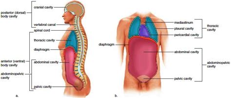
Table of Contents
The Abdominopelvic and Thoracic Cavities: Subdivisions of the Ventral Body Cavity
The human body is a marvel of intricate organization, with various systems working in harmony to maintain life. A key aspect of this organization is the division of the body into cavities, providing protection and structural support for vital organs. Two major cavities, the ventral and dorsal cavities, house the majority of the body's organs. This article focuses on the subdivisions of the ventral body cavity, specifically the thoracic cavity and the abdominopelvic cavity, exploring their contents, boundaries, and clinical significance.
The Ventral Body Cavity: A Protective Housing
The ventral body cavity, also known as the coelom, is a large, fluid-filled space that houses the majority of the internal organs. Its development is crucial for the proper formation and functioning of these organs. Unlike the dorsal cavity (which houses the brain and spinal cord), the ventral cavity is characterized by its more flexible and expandable nature, allowing for changes in organ size and position during various physiological processes like digestion and respiration. The ventral cavity is further divided into two main cavities: the thoracic cavity and the abdominopelvic cavity, separated by the diaphragm, a dome-shaped muscle essential for breathing.
The Thoracic Cavity: The Heart and Lungs' Fortress
The thoracic cavity, or chest cavity, is the superior subdivision of the ventral cavity. It’s a relatively rigid compartment primarily protected by the rib cage, sternum, and associated muscles. This robust enclosure shields the delicate organs within from external trauma. The thoracic cavity contains three major compartments:
1. The Pleural Cavities: Encasing the Lungs
Each lung resides within its own pleural cavity, a potential space lined by a serous membrane called the pleura. The pleura consists of two layers: the visceral pleura, which adheres directly to the lung surface, and the parietal pleura, which lines the inner surface of the thoracic wall. The small space between these layers contains a lubricating fluid that minimizes friction during breathing. This fluid allows the lungs to expand and contract smoothly within the chest cavity without causing damage or pain. Conditions like pneumothorax (collapsed lung), where air enters the pleural cavity, disrupt this delicate balance.
2. The Pericardial Cavity: Protecting the Heart
The pericardial cavity is a smaller, fluid-filled sac located within the mediastinum. It encloses the heart, providing protection and lubrication similar to the pleural cavities. The pericardial cavity is also lined by a serous membrane, the pericardium, with visceral and parietal layers. The pericardial fluid minimizes friction during heart contractions. Diseases such as pericarditis, an inflammation of the pericardium, can compromise this protective mechanism and lead to severe heart problems.
3. The Mediastinum: A Central Compartment
The mediastinum is a central compartment of the thoracic cavity that separates the two pleural cavities. It contains several important structures, including:
- The heart: The central pumping organ of the circulatory system.
- The great vessels: Major blood vessels like the aorta, superior and inferior vena cava, and pulmonary arteries and veins.
- The trachea: The airway that carries air to and from the lungs.
- The esophagus: The tube that transports food from the pharynx to the stomach.
- The thymus: An immune organ involved in T-cell maturation (primarily during childhood).
The Abdominopelvic Cavity: A Hub of Digestion and Reproduction
The abdominopelvic cavity is the inferior subdivision of the ventral body cavity, extending from the diaphragm to the pelvic floor. It’s further divided into two regions: the abdominal cavity and the pelvic cavity. Unlike the thoracic cavity, the abdominopelvic cavity offers less rigid protection, allowing for greater expansion and contraction related to digestive processes and pregnancy.
1. The Abdominal Cavity: Housing Digestive Organs
The abdominal cavity houses many of the major digestive organs, including:
- The stomach: Where food is churned and mixed with digestive juices.
- The small intestine: The primary site of nutrient absorption.
- The large intestine: Where water is absorbed and feces are formed.
- The liver: A vital organ involved in metabolism, detoxification, and bile production.
- The gallbladder: Stores and concentrates bile.
- The pancreas: Produces digestive enzymes and hormones like insulin and glucagon.
- The spleen: Part of the lymphatic system, involved in filtering blood and immune responses.
- The kidneys: Filter waste products from the blood and produce urine.
The abdominal cavity is lined by the peritoneum, a serous membrane similar to the pleura and pericardium. The peritoneum consists of a parietal layer lining the abdominal wall and a visceral layer covering the abdominal organs. The space between these layers, the peritoneal cavity, contains a small amount of fluid that lubricates the organs and reduces friction. Peritonitis, an inflammation of the peritoneum, can be a life-threatening condition.
2. The Pelvic Cavity: Protecting Reproductive and Urinary Organs
The pelvic cavity is the inferior portion of the abdominopelvic cavity, enclosed by the bony pelvis. It houses several vital organs, including:
- The urinary bladder: Stores urine before elimination.
- The urethra: The tube that carries urine from the bladder to the outside.
- The internal reproductive organs: In females, this includes the uterus, ovaries, and fallopian tubes. In males, this includes the prostate gland, seminal vesicles, and parts of the vas deferens.
- The rectum: The final section of the large intestine, where feces are stored before elimination.
The pelvic cavity provides protection for these sensitive organs. However, its relatively open structure means these organs are also more susceptible to injury compared to those within the thoracic cavity.
Clinical Significance: Understanding the Cavities' Role in Disease
Understanding the anatomy of the thoracic and abdominopelvic cavities is crucial in clinical practice. Many medical conditions directly relate to these cavities and their contents. For example:
- Pneumonia: Infection of the lungs within the pleural cavities.
- Pleural effusion: Accumulation of fluid in the pleural cavity.
- Pneumothorax: Air in the pleural cavity causing lung collapse.
- Pericarditis: Inflammation of the pericardium, potentially impacting heart function.
- Appendicitis: Inflammation of the appendix, located in the abdominal cavity.
- Peritonitis: Inflammation of the peritoneum, often caused by infection.
- Hepatitis: Inflammation of the liver, affecting metabolic functions.
- Pancreatitis: Inflammation of the pancreas, leading to digestive and hormonal imbalances.
- Cystitis: Inflammation of the urinary bladder in the pelvic cavity.
- Pelvic inflammatory disease (PID): Infection of the female reproductive organs in the pelvic cavity.
Diagnosing and treating these conditions often involve techniques like chest X-rays, CT scans, ultrasounds, and laparoscopic surgeries, which rely on a thorough understanding of the anatomical relationships within the thoracic and abdominopelvic cavities.
Abdominopelvic Regions and Quadrants: A Practical Division
To further facilitate the location and description of organs and pathology, the abdominopelvic cavity is often divided into nine regions or four quadrants.
Nine Abdominopelvic Regions: A Detailed Approach
This approach divides the abdominopelvic cavity into nine regions using two horizontal and two vertical planes. These regions are:
- Right hypochondriac region: Upper right region, containing the liver, gallbladder, and parts of the intestines.
- Epigastric region: Central upper region, encompassing the stomach and liver.
- Left hypochondriac region: Upper left region, containing the spleen, stomach, and parts of the intestines.
- Right lumbar region: Mid-right region, containing portions of the intestines and kidneys.
- Umbilical region: Central middle region, encompassing the umbilicus (belly button) and portions of the intestines.
- Left lumbar region: Mid-left region, containing portions of the intestines and kidneys.
- Right iliac (inguinal) region: Lower right region, containing the appendix and parts of the intestines.
- Hypogastric (pubic) region: Central lower region, containing the urinary bladder and parts of the intestines.
- Left iliac (inguinal) region: Lower left region, containing portions of the intestines.
Four Abdominopelvic Quadrants: A Simpler Approach
The simpler quadrant approach divides the abdominopelvic cavity into four sections using a single vertical and horizontal plane intersecting at the umbilicus. These quadrants are:
- Right upper quadrant (RUQ): Contains the liver, gallbladder, portions of the stomach, intestines, and pancreas.
- Left upper quadrant (LUQ): Contains the spleen, stomach, portions of the intestines, and pancreas.
- Right lower quadrant (RLQ): Contains the appendix, portions of the intestines, and reproductive organs.
- Left lower quadrant (LLQ): Contains portions of the intestines, reproductive organs, and the urinary bladder.
Both the nine-region and four-quadrant methods aid in precise anatomical location descriptions crucial for medical diagnosis and treatment.
Conclusion: Understanding the Body's Architecture
The thoracic and abdominopelvic cavities are essential components of the human body's architecture, providing vital protection and support for numerous organs. Their detailed anatomy, including the specific structures within each compartment, the serous membranes, and the regional subdivisions, is fundamental to understanding human physiology and pathology. This knowledge is crucial for medical professionals in diagnosing and treating a wide range of conditions affecting the internal organs. By appreciating the intricate organization of these cavities, we gain a deeper understanding of the remarkable complexity and resilience of the human body.
Latest Posts
Latest Posts
-
El Preterito De Los Verbos Regulares
Mar 25, 2025
-
Do Transition Metals Have A Charge
Mar 25, 2025
-
How To Calculate The Potential Difference
Mar 25, 2025
-
Why Race Is A Social Construct
Mar 25, 2025
-
Why Is Prophase The Longest Phase
Mar 25, 2025
Related Post
Thank you for visiting our website which covers about The Abdominopelvic And Thoracic Cavities Are Subdivisions Of The . We hope the information provided has been useful to you. Feel free to contact us if you have any questions or need further assistance. See you next time and don't miss to bookmark.
