The Atrioventricular Av Valves Are Closed
Muz Play
Apr 02, 2025 · 6 min read

Table of Contents
The Atrioventricular (AV) Valves are Closed: A Deep Dive into Cardiac Mechanics
The human heart, a tireless powerhouse, rhythmically pumps blood throughout our bodies. This intricate process relies on a complex interplay of chambers, vessels, and, crucially, valves. Understanding the mechanics of these valves is paramount to comprehending cardiovascular health and disease. This article delves into the specific moment when the atrioventricular (AV) valves – the mitral and tricuspid valves – are closed, exploring the physiological processes, underlying mechanisms, and clinical implications of this crucial phase of the cardiac cycle.
The Cardiac Cycle: A Stage-by-Stage Breakdown
Before focusing on the closure of the AV valves, let's establish a firm understanding of the cardiac cycle itself. This cyclical process involves two main phases: diastole and systole.
Diastole: Relaxation and Filling
Diastole represents the relaxation phase of the cardiac cycle. During this period, the heart chambers relax, allowing the ventricles to fill with blood returning from the body (through the vena cavae into the right atrium) and the lungs (through the pulmonary veins into the left atrium). Crucially, during diastole, the AV valves are OPEN, allowing passive filling of the ventricles. This passive filling is augmented by atrial contraction towards the end of diastole, which actively pushes the remaining blood into the ventricles. This stage is often characterized by a relatively low pressure within the ventricles.
Systole: Contraction and Ejection
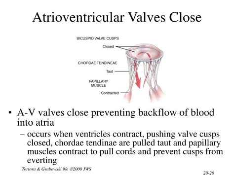
Systole is the contraction phase. This is where the powerful muscles of the ventricles contract, forcefully ejecting blood into the pulmonary artery (from the right ventricle) and the aorta (from the left ventricle). This forceful contraction significantly increases the pressure within the ventricles. It's during systole that the AV valves CLOSE, preventing the backflow of blood into the atria. This closure is essential to maintain unidirectional blood flow.
The Mechanism of AV Valve Closure: A Detailed Look
The closure of the AV valves is a passive process, driven primarily by the pressure changes within the heart chambers. Let's analyze the events leading to and the mechanics of this closure:
1. Ventricular Contraction Begins: The Pressure Gradient Shift
As ventricular systole begins, the ventricular muscles contract, causing a rapid increase in intraventricular pressure. This is the pivotal moment. Simultaneously, atrial pressure remains relatively low. This creates a crucial pressure gradient: the pressure in the ventricles now exceeds the pressure in the atria.
2. Pressure Overcomes the Valve's Resistance: The Closing Action
This newly established pressure gradient forces the AV valve leaflets (cusps) together. The increased pressure pushes the leaflets upwards, closing the valve orifice and preventing regurgitation. The papillary muscles and chordae tendineae (tendinous cords), though not actively involved in the initial closure, play a crucial role in preventing prolapse (inversion) of the valve leaflets, ensuring a tight seal.
3. The Role of Papillary Muscles and Chordae Tendineae: Preventing Prolapse
The papillary muscles are small, cone-shaped muscles within the ventricles. They are connected to the valve leaflets via the chordae tendineae. While not actively participating in valve closure, their contraction during systole helps to stabilize the valve leaflets, preventing them from inverting or prolapsing into the atria under the high ventricular pressure. This prevents regurgitation, maintaining the efficiency of the pump. Imagine them acting as a safety net, securing the valves during the forceful contraction.
4. Isovolumetric Contraction: A Brief Pause
After the AV valves close, there's a brief period known as isovolumetric contraction. During this short phase, all four heart valves are momentarily closed. The ventricles continue to contract, further increasing the pressure inside, but the volume of blood remains unchanged because no blood can enter or leave the ventricles. This builds up the pressure necessary for ventricular ejection.
Clinical Significance of AV Valve Closure Dysfunction
The efficient closure of the AV valves is crucial for maintaining optimal cardiac function. Dysfunction in this process can lead to several serious conditions:
1. Mitral Regurgitation and Tricuspid Regurgitation
If the mitral (left AV) or tricuspid (right AV) valves fail to close completely during systole, blood can leak back into the atria. This is known as mitral regurgitation or tricuspid regurgitation, respectively. This regurgitation reduces the amount of blood ejected from the ventricles, diminishing cardiac output and potentially leading to heart failure. Several factors, including valve leaflet damage, chordae tendineae rupture, and papillary muscle dysfunction, can contribute to this condition.
2. Mitral Valve Prolapse
Mitral valve prolapse occurs when one or both leaflets of the mitral valve bulge (prolapse) into the left atrium during ventricular systole. While some individuals with mitral valve prolapse are asymptomatic, it can cause regurgitation and, in severe cases, heart failure.
3. Atrial Fibrillation and Other Arrhythmias
Abnormal heart rhythms, particularly atrial fibrillation, can affect the timing and effectiveness of AV valve closure. Rapid and irregular atrial contractions can disrupt ventricular filling, causing inefficient closure and potentially contributing to regurgitation.
4. Heart Murmurs
Incomplete closure of the AV valves results in a characteristic "swishing" or "gurgling" sound – a heart murmur – which can be detected through auscultation (listening with a stethoscope). The location, timing, and characteristics of the murmur can often provide clues about the specific valve involved and the nature of the dysfunction.
Diagnostic Techniques for AV Valve Dysfunction
Several diagnostic techniques can help identify problems with AV valve closure:
- Echocardiography: This non-invasive imaging technique uses ultrasound waves to visualize the heart's structure and function, allowing clinicians to assess valve movement and identify regurgitation.
- Electrocardiography (ECG): An ECG measures the electrical activity of the heart, which can reveal irregularities in rhythm that might be associated with AV valve dysfunction.
- Cardiac Catheterization: This more invasive procedure involves inserting a thin catheter into a blood vessel to directly measure pressures within the heart chambers and assess valve function.
Management and Treatment of AV Valve Disorders
Management and treatment strategies for AV valve disorders depend on the severity of the condition and its impact on the patient's health. Options include:
- Medication: Medications, such as diuretics and ACE inhibitors, can be used to manage symptoms of heart failure associated with AV valve regurgitation.
- Surgical Intervention: In severe cases, surgical repair or replacement of the affected valve may be necessary to restore normal cardiac function. Surgical techniques have advanced significantly, offering minimally invasive approaches and improved long-term outcomes.
Conclusion: The Importance of AV Valve Function
The precise and timely closure of the atrioventricular valves is fundamental to the efficient functioning of the heart. Understanding the physiological mechanisms underlying this process, along with the potential consequences of dysfunction, is critical for clinicians and researchers alike. Advanced diagnostic techniques and improved surgical interventions have significantly improved outcomes for individuals affected by AV valve disorders, highlighting the ongoing progress in cardiovascular medicine. Further research will continue to unravel the intricacies of cardiac mechanics and lead to even more effective strategies for the prevention and treatment of these crucial valvular functions. The continuing focus on improving our understanding of the cardiac cycle and its intricacies underscores the importance of maintaining a healthy cardiovascular system for overall well-being.
Latest Posts
Latest Posts
-
Does Sohcahtoa Work On Non Right Triangles
Apr 03, 2025
-
What Are The Four Main Types Of Context
Apr 03, 2025
-
How To Know If A Graph Is Symmetric
Apr 03, 2025
-
Is Blood Clotting Negative Or Positive Feedback
Apr 03, 2025
-
Evaluating Functions Linear And Quadratic Or Cubic
Apr 03, 2025
Related Post
Thank you for visiting our website which covers about The Atrioventricular Av Valves Are Closed . We hope the information provided has been useful to you. Feel free to contact us if you have any questions or need further assistance. See you next time and don't miss to bookmark.
