The Dorsal Body Cavity Is Subdivided Into The
Muz Play
Mar 20, 2025 · 6 min read
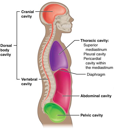
Table of Contents
The Dorsal Body Cavity: A Subdivision into Cranial and Vertebral Cavities
The human body, a marvel of intricate design, houses a complex network of organs and systems working in perfect harmony. Understanding the organization of these systems is crucial to grasping the overall function of the body. A key aspect of this organization involves the body cavities, spaces that enclose and protect vital organs. This article delves into the dorsal body cavity, exploring its subdivision and the critical structures it contains. We will examine its components, their anatomical relationships, and their clinical significance.
The Dorsal Body Cavity: A Protective Shield
The human body is broadly divided into two major cavities: the ventral body cavity and the dorsal body cavity. Unlike the ventral cavity, which houses visceral organs like the lungs and intestines, the dorsal body cavity primarily protects the central nervous system – the body's control center. This crucial role dictates its robust construction and specialized features. The dorsal body cavity is further subdivided into two distinct regions: the cranial cavity and the vertebral cavity.
The Cranial Cavity: Protecting the Brain
The cranial cavity, located within the skull, is the superior portion of the dorsal body cavity. Its primary function is to securely enclose and protect the brain, the body's master control center. The cranial bones, fused together by sutures, form a rigid and nearly impenetrable barrier against external forces. This bony protection is essential to safeguard the brain's delicate neural tissue from trauma. The cranial cavity isn't just a simple bony box; it's intricately shaped to accommodate the brain's various lobes and structures.
Key features of the cranial cavity include:
- Bony Structure: The strong, fused bones of the skull provide robust protection. These bones include the frontal bone, parietal bones, temporal bones, occipital bone, sphenoid bone, and ethmoid bone. The sutures, or joints between these bones, are immovable in adults, providing superior stability.
- Meninges: The brain is not directly in contact with the bony interior of the cranial cavity. Instead, it's enveloped by three protective membranes called the meninges: the dura mater, arachnoid mater, and pia mater. These layers provide cushioning and support, further reducing the risk of damage to the delicate brain tissue. The space between the meninges contains cerebrospinal fluid (CSF), a clear fluid that acts as a shock absorber and provides nutrients to the brain.
- Cranial Nerves: Twelve pairs of cranial nerves emerge from the brain and pass through foramina (openings) in the cranial bones. These nerves control various functions, including vision, hearing, taste, smell, and facial movements. Their carefully defined pathways through the skull ensure their protection and efficient function.
- Blood Vessels: The brain receives a rich blood supply via the internal carotid arteries and the vertebral arteries. These vessels provide essential oxygen and nutrients to the brain, ensuring its continuous activity.
The Vertebral Cavity: Protecting the Spinal Cord
The vertebral cavity, also known as the spinal canal, runs inferiorly from the cranial cavity, following the curvature of the vertebral column. This cavity is formed by the vertebral foramina, the openings in the individual vertebrae that stack upon each other. The primary function of the vertebral cavity is to protect the spinal cord, the long, cylindrical structure that extends from the brainstem to the lumbar region. The spinal cord is the central communication pathway between the brain and the rest of the body, transmitting signals to and from various organs and tissues.
Key features of the vertebral cavity:
- Vertebrae: The vertebral column is composed of 33 vertebrae, which are separated by intervertebral discs. These discs act as shock absorbers, protecting the spinal cord from jarring movements. The vertebrae's interlocking structure provides substantial strength and support.
- Spinal Cord: The spinal cord, a crucial part of the central nervous system, runs within the vertebral canal. It's protected by the bony vertebrae, intervertebral discs, and the meninges, which are continuous with those surrounding the brain. The spinal cord is responsible for transmitting nerve impulses between the brain and the peripheral nervous system.
- Spinal Nerves: Pairs of spinal nerves branch off from the spinal cord, emerging through intervertebral foramina. These nerves innervate the muscles, skin, and organs of the body. The precise location of these foramina ensures the nerves are adequately protected and can reach their target locations effectively.
- Meninges: Similar to the cranial cavity, the spinal cord is protected by three layers of meninges: the dura mater, arachnoid mater, and pia mater. The subarachnoid space, between the arachnoid and pia mater, contains cerebrospinal fluid (CSF), which cushions and nourishes the spinal cord.
Clinical Significance of the Dorsal Body Cavity
Understanding the anatomy of the dorsal body cavity is crucial in various clinical settings. Damage to structures within this cavity can have devastating consequences.
Cranial Cavity:
- Traumatic Brain Injury (TBI): Head injuries can result in fractures to the cranial bones, leading to intracranial hemorrhages (bleeding within the brain) or damage to brain tissue. The severity of TBI ranges from mild concussions to severe, life-threatening injuries.
- Meningitis: Infection of the meninges, often caused by bacteria or viruses, can cause inflammation and swelling, leading to severe headaches, fever, and even death.
- Brain Tumors: Tumors within the cranial cavity can compress brain tissue, causing neurological deficits depending on the tumor's location and size.
- Hydrocephalus: This condition involves an accumulation of cerebrospinal fluid (CSF) within the cranial cavity, causing increased intracranial pressure and potentially brain damage.
Vertebral Cavity:
- Spinal Cord Injuries: Trauma to the spine can lead to compression or severance of the spinal cord, resulting in paralysis or other neurological deficits. The severity of the injury depends on the level of the spinal cord affected.
- Spinal Stenosis: Narrowing of the vertebral canal can compress the spinal cord or nerves, causing pain, numbness, and weakness.
- Herniated Discs: Protrusion of the intervertebral discs can compress spinal nerves, causing pain, numbness, and weakness in the affected area.
- Spinal Infections: Infections such as meningitis or abscesses can affect the spinal cord and its coverings, leading to inflammation, pain, and neurological deficits.
Interconnectedness and Clinical Implications
The cranial and vertebral cavities, while distinct, are intimately connected through the continuous spinal cord. This interconnectedness has significant clinical implications. For instance, a fracture of the cervical vertebrae can not only damage the spinal cord at the site of injury but can also cause damage to the brainstem, leading to potentially fatal consequences. Similarly, conditions affecting the flow of cerebrospinal fluid in the cranial cavity can impact the spinal cord.
Conclusion
The dorsal body cavity, comprising the cranial and vertebral cavities, plays a critical role in protecting the central nervous system. The robust bony structures, protective membranes, and specialized fluids within these cavities ensure the safety and proper function of the brain and spinal cord. Understanding the anatomy and clinical significance of this cavity is essential for healthcare professionals to diagnose and treat a wide range of neurological conditions. Further research continues to unveil the complexities of this vital area, leading to improved understanding and better patient care. The detailed anatomical knowledge presented here provides a foundational understanding for further exploration in neuroanatomy and related medical fields. The importance of safeguarding this critical area for overall human health cannot be overstated.
Latest Posts
Latest Posts
-
Is Burning A Chemical Or Physical Change
Mar 21, 2025
-
How Do You Go From Liters To Moles
Mar 21, 2025
-
Heat Transfer Throught The Collision Of Moluces Direct Contacr
Mar 21, 2025
-
What Color Is The Frog Galbladder
Mar 21, 2025
-
Write The Inequality In Interval Notation
Mar 21, 2025
Related Post
Thank you for visiting our website which covers about The Dorsal Body Cavity Is Subdivided Into The . We hope the information provided has been useful to you. Feel free to contact us if you have any questions or need further assistance. See you next time and don't miss to bookmark.
