The Gram Stain Acid-fast Stain And Endospore Stain
Muz Play
Mar 17, 2025 · 6 min read
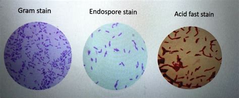
Table of Contents
The Gram Stain, Acid-Fast Stain, and Endospore Stain: Essential Differential Staining Techniques in Microbiology
Differential staining techniques are crucial tools in microbiology, allowing us to distinguish between different types of bacteria based on their unique cell wall compositions and other structural features. Among the most widely used and essential differential stains are the Gram stain, the acid-fast stain, and the endospore stain. Understanding these techniques is fundamental to accurate bacterial identification and diagnosis. This comprehensive article will delve into the principles, procedures, and significance of each stain, highlighting their applications in clinical microbiology and beyond.
The Gram Stain: A Cornerstone of Bacterial Identification
The Gram stain, developed by Hans Christian Gram in 1884, remains a cornerstone of bacterial identification in clinical microbiology laboratories worldwide. Its simplicity, speed, and diagnostic power make it an indispensable tool for differentiating bacteria into two major groups: Gram-positive and Gram-negative. This differentiation is based on fundamental differences in the bacterial cell wall structure.
Understanding Gram-Positive and Gram-Negative Cell Walls
Gram-positive bacteria possess a thick peptidoglycan layer (up to 80% of their cell wall) which retains the crystal violet-iodine complex during the decolorization step of the Gram stain. This thick peptidoglycan layer is responsible for their characteristic purple color after Gram staining. They also lack an outer membrane.
Gram-negative bacteria, on the other hand, have a thinner peptidoglycan layer (only 10-20% of their cell wall) sandwiched between an inner cytoplasmic membrane and an outer membrane containing lipopolysaccharide (LPS). The outer membrane prevents the crystal violet-iodine complex from being retained during decolorization, leading to a pink or red color after counterstaining with safranin.
The Gram Stain Procedure: A Step-by-Step Guide
The Gram stain procedure involves four key steps:
- Primary Stain (Crystal Violet): A crystal violet solution is applied to the bacterial smear, staining all cells purple.
- Mordant (Gram's Iodine): Gram's iodine acts as a mordant, forming a complex with the crystal violet within the peptidoglycan layer. This complex is larger and less likely to be removed from the cell.
- Decolorizer (Alcohol or Acetone-Alcohol): This is the crucial step differentiating Gram-positive and Gram-negative bacteria. The decolorizer dissolves the outer membrane of Gram-negative bacteria, allowing the crystal violet-iodine complex to wash away. The thick peptidoglycan layer of Gram-positive bacteria, however, resists decolorization, retaining the purple stain.
- Counterstain (Safranin): Safranin, a pink dye, is applied to stain the decolorized Gram-negative bacteria.
Significance and Applications of the Gram Stain
The Gram stain provides rapid and valuable information for guiding initial treatment decisions in bacterial infections. Knowing whether a bacterium is Gram-positive or Gram-negative helps clinicians predict its potential antibiotic susceptibility. For instance, Gram-positive bacteria are often more susceptible to penicillin and related antibiotics, while Gram-negative bacteria may require different antibiotic classes to effectively target their cell wall and other features.
The Acid-Fast Stain: Identifying Mycobacteria and Other Acid-Fast Organisms
The acid-fast stain is a differential stain specifically designed to identify bacteria with a high lipid content in their cell walls, notably members of the genus Mycobacterium, which includes the causative agents of tuberculosis and leprosy. These bacteria contain mycolic acids, a type of waxy lipid that makes them resistant to conventional staining methods, including the Gram stain.
Mycolic Acids and Acid-Fastness
Mycolic acids are long-chain fatty acids that form a complex with peptidoglycan in the cell wall of acid-fast bacteria. This waxy layer provides resistance to decolorization with acid-alcohol, a crucial characteristic in the acid-fast staining procedure.
The Acid-Fast Stain Procedure: Ziehl-Neelsen and Kinyoun Methods
Two common methods are used for acid-fast staining: the Ziehl-Neelsen method and the Kinyoun method. Both involve the application of a primary stain (carbol fuchsin), which penetrates the waxy cell wall with the aid of heat (Ziehl-Neelsen) or a modified formulation (Kinyoun). After staining, acid-alcohol is used as a decolorizer. Acid-fast bacteria retain the carbol fuchsin, while non-acid-fast bacteria are decolorized. A counterstain, such as methylene blue, is then applied to stain the decolorized bacteria.
Significance and Applications of the Acid-Fast Stain
The acid-fast stain is invaluable in the diagnosis of tuberculosis and leprosy, allowing for rapid identification of the causative bacteria in clinical specimens. Early detection and diagnosis of these diseases are crucial for effective treatment and prevention of their spread. The stain also has applications in identifying other acid-fast organisms, although these are less common in clinical settings.
The Endospore Stain: Visualizing Bacterial Endospores
Bacterial endospores are dormant, highly resistant structures formed by certain species of bacteria, primarily those belonging to the genera Bacillus and Clostridium. These spores are remarkably resistant to heat, desiccation, radiation, and chemical agents. The endospore stain is a differential stain used to visualize these structures within bacterial cells.
The Structure and Function of Endospores
Endospores are formed within the vegetative cell (the actively growing bacterial cell) as a survival mechanism under harsh environmental conditions. They contain a copy of the bacterial chromosome, essential enzymes, and a protective coat rich in dipicolinic acid and calcium ions. When conditions become favorable, the endospore germinates, giving rise to a new vegetative cell.
The Endospore Stain Procedure: Schaeffer-Fulton Method
The most commonly used method for endospore staining is the Schaeffer-Fulton method. This method utilizes malachite green, a primary stain that penetrates the spore coat with the aid of heat. After staining, water is used to decolorize the vegetative cells, while the endospores retain the malachite green. A counterstain, such as safranin, is then applied to stain the vegetative cells pink or red.
Significance and Applications of the Endospore Stain
The endospore stain is particularly useful in identifying bacterial species capable of forming endospores, which are often associated with food spoilage and various diseases. The ability to detect endospores is crucial in ensuring the sterility of medical equipment and food products, as these structures are highly resistant to sterilization methods. The stain also has applications in environmental microbiology, helping to identify spore-forming bacteria in soil and other environments.
Conclusion: The Power of Differential Staining in Microbiology
The Gram stain, acid-fast stain, and endospore stain are fundamental differential staining techniques used in microbiology to identify and characterize bacteria based on their unique cell wall structures and other cellular features. Each stain plays a crucial role in clinical diagnostics, guiding treatment decisions and preventing the spread of infectious diseases. Understanding the principles and procedures of these techniques is essential for all microbiologists and anyone working in related fields. Their continued use underscores their enduring value in the ever-evolving field of microbiology. Furthermore, the development and refinement of these staining methods have profoundly impacted our understanding of bacterial diversity and pathogenesis, making them invaluable tools for scientific research and public health efforts. Mastering these techniques opens the door to a deeper understanding of the microbial world and its impact on human health and the environment.
Latest Posts
Latest Posts
-
Which Process Takes Place In Chloroplasts
Mar 18, 2025
-
How To Do A Slope Field
Mar 18, 2025
-
How Many Electrons Can The D Orbital Hold
Mar 18, 2025
-
What Type Of Ion Do Metals Form
Mar 18, 2025
Related Post
Thank you for visiting our website which covers about The Gram Stain Acid-fast Stain And Endospore Stain . We hope the information provided has been useful to you. Feel free to contact us if you have any questions or need further assistance. See you next time and don't miss to bookmark.
