Valves That Prevent Backflow Of Blood Into The Ventricles
Muz Play
Apr 05, 2025 · 6 min read

Table of Contents
Valves That Prevent Backflow of Blood into the Ventricles: A Deep Dive into Atrioventricular and Semilunar Valves
The human heart, a tireless engine driving life's processes, relies on a sophisticated system of valves to ensure unidirectional blood flow. These valves, meticulously designed and strategically placed, prevent the backflow of blood, a critical function maintaining efficient circulation. This article delves into the fascinating world of these valves, focusing specifically on those preventing backflow into the ventricles: the atrioventricular (AV) valves and the semilunar valves. We will explore their anatomy, function, associated pathologies, and the crucial role they play in maintaining cardiovascular health.
Understanding the Heart's Chambers and Blood Flow
Before diving into the specifics of the valves, it's essential to understand the basic anatomy of the heart and the pathway of blood flow. The heart consists of four chambers: two atria (upper chambers) and two ventricles (lower chambers). Deoxygenated blood enters the right atrium from the body via the superior and inferior vena cava. It then flows into the right ventricle, which pumps it to the lungs for oxygenation via the pulmonary artery. Oxygenated blood returns from the lungs to the left atrium via the pulmonary veins. Finally, it passes into the left ventricle, which pumps it out to the body through the aorta.
This intricate dance of blood flow is orchestrated by the precise opening and closing of the heart valves. These valves ensure that blood moves in one direction only, preventing any regurgitation or backflow. This prevents inefficient circulation and protects the heart from overworking.
The Atrioventricular (AV) Valves: Guardians of Ventricular Filling
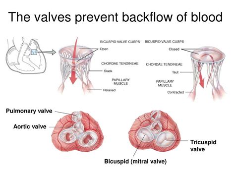
The atrioventricular valves, positioned between the atria and the ventricles, prevent backflow from the ventricles into the atria during ventricular contraction (systole). There are two AV valves:
1. The Tricuspid Valve: Right Atrium to Right Ventricle
Located between the right atrium and the right ventricle, the tricuspid valve is aptly named for its three cusps (leaflets) of fibrous tissue. These cusps are connected to papillary muscles via chordae tendineae, strong tendinous cords resembling tiny strings. The papillary muscles contract simultaneously with the ventricular walls, preventing the cusps from inverting into the atrium during ventricular contraction. This coordinated action ensures that blood flows smoothly from the right atrium to the right ventricle and prevents regurgitation. Dysfunction in the tricuspid valve can lead to tricuspid regurgitation, a condition where blood flows back into the right atrium.
2. The Mitral Valve (Bicuspid Valve): Left Atrium to Left Ventricle
The mitral valve, also known as the bicuspid valve due to its two cusps, sits between the left atrium and the left ventricle. Similar to the tricuspid valve, it is anchored to papillary muscles via chordae tendineae, ensuring its proper functioning. The mitral valve handles the higher pressures associated with the systemic circulation, making it susceptible to certain pathologies. Mitral valve prolapse, where one or both cusps bulge back into the left atrium during systole, is a common disorder. Mitral stenosis, a narrowing of the valve opening, restricts blood flow from the left atrium to the left ventricle.
The Semilunar Valves: Preventing Backflow from the Arteries
The semilunar valves, positioned at the exits of the ventricles, prevent backflow from the arteries into the ventricles during ventricular relaxation (diastole). There are two semilunar valves:
1. The Pulmonary Valve: Right Ventricle to Pulmonary Artery
The pulmonary valve is located between the right ventricle and the pulmonary artery. Unlike the AV valves, the semilunar valves don't have chordae tendineae. Instead, their three cusps are shaped like half-moons, fitting together tightly to prevent backflow. The pulmonary valve's function is to prevent blood from flowing back into the right ventricle after it's been ejected into the pulmonary artery. Pulmonary stenosis, a narrowing of the pulmonary valve opening, restricts blood flow to the lungs. Pulmonary regurgitation, a backflow of blood from the pulmonary artery into the right ventricle, can also occur.
2. The Aortic Valve: Left Ventricle to Aorta
The aortic valve sits between the left ventricle and the aorta, the largest artery in the body. This valve, with its three semilunar cusps, prevents the backflow of oxygenated blood from the aorta into the left ventricle during diastole. The aortic valve handles the highest pressures within the cardiovascular system, making it prone to specific pathologies. Aortic stenosis, a narrowing of the aortic valve opening, is a significant health concern. Aortic regurgitation, the backward flow of blood from the aorta into the left ventricle, can also be a serious condition.
Pathologies Affecting the Heart Valves
Valvular heart disease encompasses a range of conditions affecting the structure and function of the heart valves. These diseases can be either congenital (present at birth) or acquired (developing later in life). Some common pathologies include:
- Stenosis: Narrowing of the valve opening, restricting blood flow.
- Regurgitation (or insufficiency): Leaky valve, allowing blood to flow backward.
- Prolapse: Bulging of one or more valve cusps into the adjacent chamber.
These conditions can lead to various symptoms, including shortness of breath (dyspnea), chest pain (angina), fatigue, and palpitations. The severity of symptoms depends on the extent of valve dysfunction and the individual's overall health.
Diagnosis and Treatment of Valvular Heart Disease
Diagnosing valvular heart disease typically involves a thorough physical examination, including auscultation (listening to the heart sounds with a stethoscope) to detect murmurs (abnormal heart sounds). Further diagnostic tests may include:
- Echocardiogram: Ultrasound imaging of the heart, providing detailed images of the heart chambers and valves.
- Electrocardiogram (ECG): Measures the electrical activity of the heart.
- Cardiac catheterization: A minimally invasive procedure allowing direct visualization of the heart chambers and valves.
Treatment options for valvular heart disease vary depending on the severity of the condition and the individual's overall health. Options include:
- Medication: To manage symptoms and slow disease progression.
- Valve repair: Surgical or catheter-based procedures to repair a damaged valve.
- Valve replacement: Surgical replacement of a damaged valve with a prosthetic valve (mechanical or biological).
The Importance of Maintaining Cardiovascular Health
Maintaining good cardiovascular health is crucial in preventing or delaying the onset of valvular heart disease. This involves:
- Adopting a healthy lifestyle: Regular exercise, a balanced diet, and maintaining a healthy weight.
- Managing risk factors: Controlling blood pressure, cholesterol levels, and blood sugar.
- Quitting smoking: Smoking significantly increases the risk of cardiovascular disease.
Regular check-ups with a healthcare provider are essential for early detection and management of any cardiovascular problems.
Conclusion: The Unsung Heroes of the Circulatory System
The atrioventricular and semilunar valves are unsung heroes of the circulatory system, diligently working to ensure efficient and unidirectional blood flow through the heart. Their intricate anatomy and synchronized function are critical for maintaining cardiovascular health. Understanding their roles, associated pathologies, and available treatment options is essential for both healthcare professionals and the general public. By embracing a healthy lifestyle and seeking regular medical check-ups, we can contribute to the longevity and well-being of these vital valves and, consequently, our overall cardiovascular health. The precision and efficiency of these valves highlight the incredible complexity and beauty of the human body's design. Further research continues to refine our understanding of these crucial components of the heart, leading to improved diagnostic tools and treatment strategies for valvular heart disease.
Latest Posts
Latest Posts
-
Formulas With Polyatomic Ions Worksheet Answers
Apr 06, 2025
-
Practicing Dna Transcription And Translation Answer Key
Apr 06, 2025
-
How To Prove A Transformation Is Linear
Apr 06, 2025
-
Is The Mutant Allele Dominant Or Recessive
Apr 06, 2025
-
Why Do Covalent Compounds Have Low Melting Points
Apr 06, 2025
Related Post
Thank you for visiting our website which covers about Valves That Prevent Backflow Of Blood Into The Ventricles . We hope the information provided has been useful to you. Feel free to contact us if you have any questions or need further assistance. See you next time and don't miss to bookmark.
