What Color Is The Animal Cell
Muz Play
Mar 22, 2025 · 5 min read
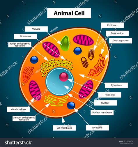
Table of Contents
- What Color Is The Animal Cell
- Table of Contents
- What Color Is an Animal Cell? The Surprising Answer and Beyond
- The Invisible World of Animal Cells: Why Color Is Relative
- The Role of Staining Techniques
- Factors Influencing Apparent Cell Color
- Beyond the Visible Spectrum: Exploring Invisible Aspects
- Advanced Imaging Techniques
- The Importance of Understanding Animal Cell Structure
- Conclusion: Color Is Just the Beginning
- Latest Posts
- Latest Posts
- Related Post
What Color Is an Animal Cell? The Surprising Answer and Beyond
The question, "What color is an animal cell?" might seem simple at first glance. However, the answer is far more nuanced than a single color. The truth is, animal cells don't have a single defining color. Their appearance under a microscope depends heavily on several factors, including staining techniques, the type of cell, and even the age and health of the cell. This article will delve into the complexities of animal cell visualization and explore the reasons behind the seemingly simple question's multifaceted answer.
The Invisible World of Animal Cells: Why Color Is Relative
Animal cells, the fundamental building blocks of animals, are typically microscopic, meaning they're too small to be seen with the naked eye. To observe them, we need the help of microscopes, often powerful ones capable of magnification at hundreds or even thousands of times. Without any staining or specialized preparation, these cells appear nearly transparent or colorless under a light microscope. This is because their constituent parts – the cytoplasm, nucleus, and organelles – are largely composed of water and other colorless substances. Imagine looking at a drop of clear water – you wouldn't see much detail unless something alters its transparency.
The Role of Staining Techniques
To visualize the intricate structures within animal cells, scientists employ various staining techniques. These techniques use dyes or stains that bind to specific cellular components, making them visible under the microscope. The color observed thus becomes entirely dependent on the specific stain used.
-
Hematoxylin and Eosin (H&E) Staining: This is a very common stain used in histology (the study of tissues). Hematoxylin stains the cell nucleus a deep purple or blue, while eosin stains the cytoplasm a pinkish-red. This combination provides excellent contrast, allowing for the identification of different cell types and structures within tissues. Using H&E, you'd see a mix of purplish-blue and pinkish-red, far from a single color.
-
Other Common Stains: Many other stains target specific organelles or molecules within the cell. For example, some stains might highlight the green fluorescence of certain proteins, or others might reveal the brown presence of lipids. The choice of stain directly determines the colors you observe.
Factors Influencing Apparent Cell Color
Beyond staining techniques, several factors contribute to the observed color of animal cells, even when viewed with stains:
-
Cell Type: Different cell types have varied compositions and structures. For example, a muscle cell might have a different concentration of proteins than a nerve cell, potentially affecting its interaction with stains and resulting in subtle color variations.
-
Cell Age and Health: Older or unhealthy cells may exhibit different staining patterns than younger, healthy cells. Changes in cellular structures due to aging or disease can influence how stains bind, affecting the perceived color.
-
Preparation Techniques: The method of preparing the cells for microscopy can influence the final image. Fixation methods, which preserve the cell's structure, can alter the cell's permeability to stains, impacting the staining intensity and overall color.
-
Microscope Type: While light microscopy is commonly used, other advanced techniques, such as fluorescence microscopy or electron microscopy, reveal different aspects of the cell's structure. Fluorescence microscopy, for instance, uses fluorescent dyes, generating images in a range of colors depending on the dye used. Electron microscopy, on the other hand, doesn't rely on color but provides highly detailed images based on electron density.
Beyond the Visible Spectrum: Exploring Invisible Aspects
The visual color of an animal cell under a microscope is just one small piece of the puzzle. Much of the cell's complexity lies beyond the visible spectrum. Modern techniques allow scientists to investigate cellular processes and components invisible to the naked eye and even to standard light microscopy.
Advanced Imaging Techniques
Techniques such as:
-
Confocal Microscopy: Allows for high-resolution 3D imaging of cells by eliminating out-of-focus light. This technique can reveal intricate details of cellular structures, although colors observed are again influenced by the fluorescent probes used.
-
Super-Resolution Microscopy: Overcomes the diffraction limit of light microscopy, enabling the visualization of structures smaller than the wavelength of light. Again, colors observed are related to the fluorescent markers.
-
Electron Microscopy: Uses electrons instead of light to create images, offering unparalleled resolution but not in the context of color. Electron microscopy reveals intricate details of cellular ultrastructure, but the resulting images are typically in grayscale.
These advanced techniques provide insights into the dynamic processes within the cell, far exceeding the limitations of simply describing the cell's color. They reveal the complexity of cellular architecture, revealing the intricate networks of protein interactions, the precise positioning of organelles, and the dynamic movements of molecules within the cell.
The Importance of Understanding Animal Cell Structure
Understanding the structure and function of animal cells is fundamental to biology and medicine. This knowledge helps us understand:
-
Disease Processes: Many diseases arise from malfunctioning cells. Visualizing cells using microscopy techniques allows researchers to investigate how cells are affected by disease, helping to develop new diagnostic tools and treatments.
-
Drug Development: Understanding cellular mechanisms is crucial for developing effective drugs that target specific cellular processes. Microscopy is an essential tool for evaluating the efficacy and potential side effects of new drugs.
-
Tissue Engineering: Regenerative medicine relies on our ability to grow and manipulate cells in the lab to repair damaged tissues. Observing cell behavior and interaction is crucial to the success of this field.
-
Developmental Biology: Studying cell development is essential for understanding how organisms grow and differentiate. Microscopy allows us to track cell changes over time, revealing the intricate processes involved in growth and development.
Conclusion: Color Is Just the Beginning
The question "What color is an animal cell?" highlights the fact that the visual appearance of a cell is highly dependent on various factors, especially staining techniques and microscopy methods. While stained cells can appear in a variety of colors, from deep purple and pinkish-red to various fluorescent hues, the true beauty and complexity of animal cells lie far beyond what we can see with our eyes, or even with a basic light microscope. Advanced imaging techniques offer unprecedented insights into the dynamic processes within these remarkable units of life, underscoring the importance of studying cell structure and function for advancements in biology and medicine. The next time you consider the color of an animal cell, remember the complex world of cellular machinery that lies beneath the surface of that seemingly simple question.
Latest Posts
Latest Posts
-
A First Course In Differential Equations Book
Mar 25, 2025
-
Moment Of Intertia Of A Rod
Mar 25, 2025
-
What Determines The Function Of A Protein
Mar 25, 2025
-
What Is Explained By The Sliding Filament Theory
Mar 25, 2025
-
Polarity Lead To Heat Of Vaporization
Mar 25, 2025
Related Post
Thank you for visiting our website which covers about What Color Is The Animal Cell . We hope the information provided has been useful to you. Feel free to contact us if you have any questions or need further assistance. See you next time and don't miss to bookmark.
