What Is A Wet Mount Slide
Muz Play
Apr 03, 2025 · 6 min read

Table of Contents
What is a Wet Mount Slide? A Comprehensive Guide
A wet mount slide is a simple yet crucial technique in microscopy, providing a quick and easy way to observe living organisms and specimens in their natural, hydrated state. Unlike prepared slides with permanently mounted specimens, wet mounts allow for the examination of dynamic processes, such as movement and cellular activity, in real-time. This technique is widely employed in various fields, including biology, microbiology, and medicine, making it an essential tool for researchers and students alike. This comprehensive guide will delve into every aspect of wet mount slide preparation, from the materials required to troubleshooting common issues, ensuring you master this fundamental microscopy skill.
Understanding the Basics: Why Use a Wet Mount?
The primary advantage of a wet mount lies in its ability to observe specimens in a near-natural environment. The aqueous medium prevents the specimen from drying out, maintaining its structure and allowing for observation of natural behaviors. This is especially important for living organisms, which require a hydrated environment to survive and exhibit their typical characteristics. Key advantages include:
- Observation of Living Organisms: The wet mount allows for the study of the motility and behavior of living microorganisms like bacteria, protozoa, and algae.
- Maintaining Specimen Integrity: The aqueous medium preserves the natural shape and structure of the specimen, preventing artifacts that might occur from drying or fixation.
- Simplicity and Speed: Preparation is quick and requires minimal equipment, making it ideal for quick observations and classroom demonstrations.
- Cost-Effectiveness: The materials needed are inexpensive and readily available.
- Versatility: Applicable to a wide range of specimens, from single-celled organisms to larger tissue samples.
Essential Materials for Wet Mount Preparation
Before embarking on wet mount preparation, ensure you have gathered the necessary materials. These include:
- Microscope Slides: Standard glass microscope slides provide a stable and transparent platform for observation.
- Coverslips: Thin, square glass coverslips are used to flatten the specimen and prevent it from drying out. The size of the coverslip should be appropriate for the size of the slide and the specimen.
- Specimen: This can range from a drop of pond water teeming with microorganisms to a prepared sample of cells or tissue.
- Pipette or Dropper: Used to transfer the specimen onto the slide.
- Dissecting Needle or Forceps: Useful for manipulating the specimen, especially if it's a solid sample.
- Water or Mounting Medium: Distilled water is generally preferred, but other mounting media, like saline solution or stains, can be used depending on the specimen and the desired outcome. A mounting medium with a higher refractive index can improve clarity.
- Lens Paper or Kimwipes: Used to clean the slides and coverslips before and after use.
Step-by-Step Wet Mount Slide Preparation
Creating a successful wet mount involves a careful and methodical approach. Here's a step-by-step guide:
-
Cleaning: Begin by thoroughly cleaning the microscope slide and coverslip with lens paper or Kimwipes. Any dust or debris on the surface can interfere with observation.
-
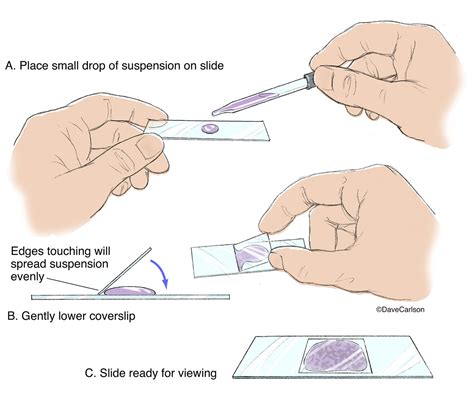
Specimen Placement: Place a small drop of the specimen onto the center of the clean microscope slide using a pipette or dropper. Avoid using an excessive amount; a single, small drop is usually sufficient. Too much liquid can overflow the coverslip and create artifacts.
-
Coverslip Application: Carefully lower the coverslip onto the slide, at a 45-degree angle, to prevent the formation of air bubbles. Slowly lower the coverslip to avoid trapping air bubbles. If air bubbles do appear, gently tap the coverslip with a pencil eraser or gently press down on the coverslip to remove them.
-
Excess Liquid Removal: If there is excess liquid, gently blot any excess with a piece of absorbent paper. You should only have a thin layer of liquid beneath the coverslip.
-
Microscope Observation: Place the slide onto the microscope stage and begin your observations. Start with the lowest magnification objective lens to locate the specimen, then gradually increase the magnification to view finer details.
Advanced Techniques and Variations
While the basic wet mount is straightforward, several variations and advanced techniques enhance its capabilities:
-
Staining: Adding stains to the specimen can highlight specific cellular structures or improve contrast. Common stains include methylene blue, iodine, and crystal violet, each providing unique visualization properties. Remember to consider the potential impact of the stain on the living specimen.
-
Hanging Drop Slide: This variation involves suspending the specimen in a drop of liquid hanging from the underside of a coverslip. It's particularly useful for observing motile organisms in a less constrained environment.
-
Using Mounting Media: Alternatives to water, such as saline solutions, glycerine, or specialized mounting media, can improve the longevity of the wet mount or enhance the visibility of certain structures. These can slow down the movement of the organisms under observation or even preserve them for longer periods.
-
Pressure Mounts: For delicate specimens, a slight pressure applied to the coverslip can help flatten and spread the sample for better viewing. However, this should be done with extreme caution to avoid damaging the specimen.
Troubleshooting Common Wet Mount Problems
Despite the simplicity of the technique, several issues can arise during wet mount preparation. Here’s how to troubleshoot common problems:
-
Air Bubbles: Air bubbles obscure the view. Gently tapping the coverslip or lowering it more carefully can usually resolve this issue.
-
Excessive Liquid: Excessive liquid can overflow and make focusing difficult. Use a small amount of liquid initially and blot away any excess.
-
Specimen Too Thick or Dense: This can make focusing difficult. Try diluting the sample with water or using a smaller amount.
-
Coverslip Movement: The coverslip should remain in place. If it moves, reapply it more carefully, ensuring a good seal.
-
Specimen Drying Out: If the specimen dries, the observation will be affected. Seal the edges of the coverslip with petroleum jelly to prevent rapid evaporation, though this is generally not necessary for short observations.
Applications of Wet Mounts Across Various Disciplines
Wet mounts are indispensable across numerous scientific fields:
-
Biology: Observing living microorganisms like bacteria, protozoa, and algae in their natural environment. Studying cell division, movement, and other dynamic processes.
-
Microbiology: Identifying bacteria and other microorganisms. Testing the effectiveness of antibiotics or other antimicrobial agents.
-
Medicine: Examining blood smears, urine samples, or other bodily fluids for the presence of pathogens or other abnormalities.
-
Parasitology: Identifying parasitic organisms in fecal samples or other clinical specimens.
-
Environmental Science: Analyzing water samples for the presence of microorganisms and assessing water quality.
-
Education: Wet mounts are an excellent tool for teaching basic microscopy techniques and demonstrating the diversity of life.
Safety Precautions When Handling Wet Mounts
While wet mount preparation is relatively safe, some precautions are necessary:
-
Handle slides and coverslips with care: Avoid scratching or breaking them.
-
Dispose of used slides and coverslips properly: Follow your institution's guidelines for waste disposal.
-
Use appropriate personal protective equipment (PPE): If working with potentially hazardous specimens, wear gloves and eye protection.
-
Clean up spills immediately: Avoid slipping hazards.
-
Always sterilize used equipment properly: Especially when handling biological samples.
Conclusion: Mastering the Wet Mount Technique
The wet mount slide is a fundamental technique in microscopy, offering a simple yet powerful method for observing living organisms and specimens in their natural state. By understanding the principles involved, mastering the preparation steps, and learning to troubleshoot common problems, you can harness the power of the wet mount to unlock the wonders of the microscopic world. Its versatility and accessibility make it an indispensable tool for scientists, educators, and anyone fascinated by the intricacies of life at a microscopic scale. Remember to always practice careful and safe procedures when preparing and handling your wet mounts.
Latest Posts
Latest Posts
-
An Unsaturated Fatty Acid Resulting From Hydrogenation Is Known As
Apr 04, 2025
-
What Are The Elements That Make Up Salt
Apr 04, 2025
-
Ionic Compound For Sodium And Sulfur
Apr 04, 2025
-
Cells Are Basic Unit Of Life
Apr 04, 2025
-
Two Bones That Form The Nasal Septum
Apr 04, 2025
Related Post
Thank you for visiting our website which covers about What Is A Wet Mount Slide . We hope the information provided has been useful to you. Feel free to contact us if you have any questions or need further assistance. See you next time and don't miss to bookmark.
