What Is Involved In Making A Smear
Muz Play
Mar 22, 2025 · 7 min read
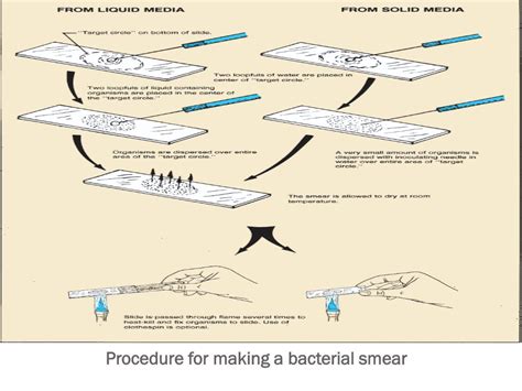
Table of Contents
What's Involved in Making a Smear: A Comprehensive Guide
Making a smear, whether for medical diagnostics or scientific research, is a crucial process requiring precision and attention to detail. This seemingly simple procedure involves several intricate steps, each impacting the final result's accuracy and reliability. This comprehensive guide delves into the complexities of smear preparation, covering various types, techniques, and essential considerations for optimal outcomes.
Understanding the Purpose of Smears
Before delving into the specifics of smear preparation, understanding the purpose is vital. Smears, also known as smears preparations, are thin layers of a specimen spread on a microscopic slide. This spreading process creates a monolayer of cells or organisms, allowing for individual examination under a microscope. Their purpose varies depending on the context:
Medical Diagnostics:
- Identifying pathogens: Smears are fundamental in diagnosing infectious diseases, allowing the identification of bacteria, fungi, parasites, and viruses. Gram staining, for example, is a crucial smear technique for differentiating bacteria based on their cell wall structure.
- Detecting abnormal cells: Pap smears, for instance, are essential in detecting precancerous and cancerous cells in the cervix. These smears help in early cancer detection and improved treatment outcomes.
- Analyzing blood cells: Blood smears enable the examination of blood cells, identifying abnormalities like anemia, leukemia, or infections. Differential blood counts rely heavily on accurate smear preparation.
Scientific Research:
- Microscopic examination of cells and tissues: Researchers extensively use smears to study cellular structures, processes, and interactions. This can range from studying microbial communities to analyzing cellular responses to specific treatments.
- Cytogenetic analysis: Smears are crucial for karyotyping, a technique for visualizing and analyzing chromosomes to detect chromosomal abnormalities.
- Immunocytochemistry: Smears are used as substrates for immunocytochemical staining, allowing researchers to locate and identify specific proteins or antigens within cells.
Types of Smears and Their Applications
Several types of smears exist, each tailored to specific applications and specimen types:
1. Direct Smears:
These are the simplest form of smears. A small amount of specimen is directly applied to a clean slide and spread thinly using a spreader slide or a loop. This technique is commonly used for examining fresh specimens, such as bacterial cultures or body fluids.
2. Fixed Smears:
Unlike direct smears, fixed smears undergo a fixation process to preserve cellular morphology and prevent degradation. Fixatives, such as methanol or heat, are used to coagulate proteins, preventing cellular distortion during staining and microscopic examination. This is especially important for long-term storage and preservation of samples.
3. Buffered Smears:
Certain specimens require a buffered environment to maintain cellular integrity and optimal staining results. These buffered smears ensure the pH remains within a specific range, preventing artifacts and enhancing staining quality.
4. Concentrated Smears:
When dealing with low-concentration specimens, concentration techniques might be necessary before smear preparation. Centrifugation is often employed to concentrate the cells or organisms, increasing the chances of detecting them during microscopic examination.
The Step-by-Step Process of Making a Smear
The actual process of making a smear involves several critical steps:
1. Preparation:
- Gather materials: Assemble all necessary materials: clean microscope slides, appropriate specimen, spreader slide or inoculation loop (depending on the smear type), fixative (if required), staining reagents (if applicable), and a microscope. Clean slides are crucial to avoid contamination and artifacts. Properly label slides with identifiers to prevent sample mix-ups.
- Specimen preparation: The preparation of the specimen varies significantly depending on the type of specimen (e.g., blood, tissue, bacterial culture). Ensure the specimen is appropriately collected, handled, and processed according to established protocols.
2. Smear Preparation:
- Direct Smear Technique: A small drop of specimen is placed near one end of the clean slide. Using a spreader slide at a 45-degree angle, gently spread the specimen across the slide in a thin, even layer. Avoid creating streaks or thick areas that hinder microscopic examination.
- Bacterial Smear Technique: For bacterial cultures, a sterile inoculation loop is used to transfer a small amount of bacteria to the slide. The bacteria are then spread evenly across the slide using the loop.
- Blood Smear Technique: A small drop of blood is placed near one end of the slide. Using a spreader slide, a thin film of blood is created by pulling the spreader slide across the drop of blood. The angle and speed control the thickness of the smear.
3. Fixation (If Applicable):
- Heat Fixation: Pass the smear gently through a Bunsen burner flame several times. This denatures proteins and adheres the cells to the slide, preventing them from washing away during staining. Overheating can distort cells, so careful execution is essential.
- Chemical Fixation: Immerse the slide in a suitable fixative, such as methanol or formalin, for a specific time period. Chemical fixation preserves cellular morphology without the risk of heat damage associated with heat fixation.
4. Staining (If Applicable):
Staining enhances the visibility of cellular structures and aids in identification. The choice of staining technique depends on the type of specimen and the information sought. Common staining techniques include:
- Gram staining: Differentiates bacteria based on their cell wall structure.
- Acid-fast staining: Identifies acid-fast bacteria, such as Mycobacterium tuberculosis.
- Giemsa staining: Used for blood smears and identifying parasites.
- Wright's stain: Another popular stain for blood smears.
5. Microscopy:
Once the smear is stained (if applicable) and dry, it's ready for microscopic examination. The appropriate objective lens should be selected, depending on the size and features of interest. Careful observation and documentation of findings are crucial for accurate interpretation.
Factors Influencing Smear Quality
Several factors can influence the quality of a smear and its subsequent analysis:
- Specimen quality: A poor-quality specimen will inherently limit the information obtainable from the smear.
- Technique: Proper technique is paramount; poor spreading can create areas too thick or thin for analysis, obscuring cellular detail.
- Fixation: Inadequate or excessive fixation can distort cellular morphology, affecting diagnosis and interpretation.
- Staining: Incorrect staining procedures can lead to artifacts and misidentification of cells or organisms.
- Microscopy skills: The microscopist's skill in identifying cellular features and recognizing artifacts is vital for accurate results.
Troubleshooting Common Issues in Smear Preparation
Several challenges can arise during smear preparation. Identifying and addressing these issues is crucial for ensuring reliable results:
- Uneven Smear: This often results from improper spreading technique. Practice is key to achieving a consistent, even smear.
- Too Thick Smear: Thick smears obstruct microscopic examination, hindering the visualization of individual cells. Use smaller specimen volumes and adjust spreading techniques.
- Too Thin Smear: Extremely thin smears may lack sufficient cells for analysis. Use larger specimen volumes and adjust spreading techniques.
- Artifacts: Artifacts, such as air bubbles or debris, can interfere with microscopic examination. Careful handling and meticulous slide preparation can minimize artifacts.
- Poor Staining: Problems with staining can stem from various factors, including expired reagents, incorrect staining times, or inadequate rinsing. Adhering to standardized staining protocols is essential.
Advanced Techniques and Considerations
For advanced applications, specialized techniques and considerations might be necessary:
- Immunocytochemistry: This technique uses antibodies to detect specific proteins within cells, enhancing the identification of specific cell types or pathogens.
- Fluorescence in situ hybridization (FISH): FISH allows for the visualization of specific DNA sequences within cells, often used in cytogenetic studies.
- Digital Microscopy: Digital microscopy provides high-resolution images and facilitates image analysis using computer software.
Conclusion
Creating a high-quality smear is a precise process with significant implications for medical diagnostics and scientific research. This comprehensive guide outlines the crucial steps involved, emphasizing the importance of technique, specimen quality, and appropriate staining methods. By understanding these intricacies and mastering the techniques, researchers and medical professionals can ensure the accuracy and reliability of smear-based analyses. Consistent practice and attention to detail are key to producing optimal smears for accurate interpretations and reliable results. Remember to always adhere to safety protocols when handling biological specimens.
Latest Posts
Latest Posts
-
In Glycolysis What Starts The Process Of Glucose Oxidation
Mar 22, 2025
-
Is Ice Melting A Chemical Change Or Physical
Mar 22, 2025
-
Most Influential Person In The 20th Century
Mar 22, 2025
-
Cell A Basic Unit Of Life
Mar 22, 2025
-
What Is The Electron Configuration Of N
Mar 22, 2025
Related Post
Thank you for visiting our website which covers about What Is Involved In Making A Smear . We hope the information provided has been useful to you. Feel free to contact us if you have any questions or need further assistance. See you next time and don't miss to bookmark.
