What Is The Function Of Atrioventricular Valves
Muz Play
Mar 30, 2025 · 6 min read
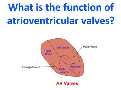
Table of Contents
What is the Function of Atrioventricular Valves? A Deep Dive into Cardiac Physiology
The human heart, a tireless engine of life, relies on a complex interplay of chambers, vessels, and valves to efficiently pump blood throughout the body. Central to this intricate system are the atrioventricular (AV) valves, crucial structures that ensure unidirectional blood flow between the heart's atria and ventricles. Understanding their function is key to grasping the fundamental mechanics of the cardiovascular system. This article will delve into the detailed anatomy, physiology, and clinical significance of the AV valves, exploring their role in maintaining circulatory health.
Anatomy of the Atrioventricular Valves
The human heart possesses two AV valves: the tricuspid valve and the mitral valve (also known as the bicuspid valve). Both are composed of leaflets (or cusps) of fibrous connective tissue, covered by endocardium, the innermost layer of the heart wall. These leaflets are connected to papillary muscles within the ventricles via chordae tendineae – strong, fibrous strings resembling tiny tendons.
The Tricuspid Valve
Located between the right atrium and the right ventricle, the tricuspid valve typically has three leaflets. Its name, derived from "tri" (three) and "cusp" (leaflets), aptly reflects its structure. This valve prevents backflow of blood from the right ventricle into the right atrium during ventricular systole (contraction).
The Mitral Valve
The mitral valve, situated between the left atrium and the left ventricle, is characterized by its two leaflets. Its alternative name, "bicuspid valve," originates from "bi" (two) and "cusp," highlighting its two-leaflet structure. Like the tricuspid valve, the mitral valve ensures that blood flows only from the left atrium to the left ventricle during ventricular systole. The mitral valve's robust construction is crucial, as it handles the higher pressures associated with the systemic circulation.
Physiology of Atrioventricular Valve Function
The AV valves' primary function is to maintain unidirectional blood flow, preventing regurgitation (backflow) during ventricular contraction. This precise control is achieved through a coordinated sequence of events during the cardiac cycle:
Diastole (Relaxation): The Valves Open
During diastole, the ventricles relax, and their pressure decreases. This lower ventricular pressure allows blood from the atria to passively flow into the ventricles, filling them in preparation for the next contraction. At this stage, the AV valves are open, allowing for this unimpeded blood flow. The chordae tendineae are relaxed, allowing the leaflets to open fully.
Systole (Contraction): The Valves Close
As the ventricles begin to contract (systole), the pressure within them rapidly increases. This increased pressure pushes the AV valve leaflets together, closing the valves. This closure prevents blood from flowing back into the atria. Simultaneously, the papillary muscles contract, tightening the chordae tendineae. This crucial step prevents the leaflets from inverting (prolapsing) into the atria under the high ventricular pressure. The coordinated action of valve leaflets, papillary muscles, and chordae tendineae ensures effective valve closure and prevents regurgitation.
The Role of Papillary Muscles and Chordae Tendineae
The papillary muscles and chordae tendineae are integral to the AV valves' proper function. They play a vital role in preventing valve prolapse, a condition where the valve leaflets bulge backward into the atria during ventricular contraction. This prolapse can lead to regurgitation, reducing the efficiency of the heart's pumping action. The coordinated contraction of the papillary muscles prevents this prolapse by maintaining tension on the chordae tendineae, securing the leaflets in their closed position during systole.
Clinical Significance of Atrioventricular Valve Dysfunction
Dysfunction of the AV valves can lead to significant cardiovascular complications. The most common problems include:
Mitral Valve Prolapse (MVP)
MVP is a condition where one or both leaflets of the mitral valve bulge back into the left atrium during ventricular systole. While many individuals with MVP are asymptomatic, it can cause mitral regurgitation, leading to shortness of breath, fatigue, and eventually heart failure.
Mitral Regurgitation (MR)
MR occurs when the mitral valve doesn't close tightly, allowing blood to leak backward from the left ventricle to the left atrium during systole. This can strain the heart, leading to symptoms similar to MVP, including shortness of breath, fatigue, and palpitations. Severe MR can lead to heart failure.
Tricuspid Regurgitation (TR)
TR, analogous to MR, involves the tricuspid valve failing to close properly, allowing blood to leak backward from the right ventricle to the right atrium during systole. While often less severe than MR, TR can contribute to heart failure and other complications.
Atrioventricular Valve Stenosis
Stenosis refers to the narrowing of a valve opening, restricting blood flow. AV valve stenosis can occur in both the mitral and tricuspid valves. This narrowing increases the workload on the heart, leading to symptoms like shortness of breath, fatigue, and chest pain (angina).
Diagnostic Methods for AV Valve Disease
Several diagnostic methods are available to assess the health and function of the AV valves:
Echocardiography
Echocardiography, a non-invasive imaging technique using ultrasound, is the primary method for evaluating AV valve function. It allows visualization of the valve structures, assesses their movement, and identifies any regurgitation or stenosis.
Cardiac Catheterization
Cardiac catheterization, a more invasive procedure, involves inserting a catheter into a blood vessel to reach the heart chambers. This allows for direct measurement of pressure gradients across the AV valves and assessment of valve function.
Electrocardiography (ECG)
ECG, which records the heart's electrical activity, can provide indirect evidence of AV valve disease. Certain ECG patterns can suggest valve dysfunction, though echocardiography is necessary for definitive diagnosis.
Treatment Options for AV Valve Disease
Treatment for AV valve disease depends on the severity of the condition and the presence of symptoms. Options include:
Medical Management
For mild cases of AV valve disease, medical management might focus on managing symptoms with medications to reduce heart strain. These medications might include diuretics to reduce fluid retention and ACE inhibitors to manage blood pressure.
Surgical Intervention
More severe cases of AV valve disease often require surgical intervention. This might involve:
- Valve repair: This procedure aims to restore the valve's proper function without replacing it. It’s generally preferred over valve replacement when feasible.
- Valve replacement: This involves replacing the damaged valve with a prosthetic valve, either mechanical or biological. The choice of valve depends on factors such as the patient's age and overall health.
Conclusion
The atrioventricular valves are indispensable components of the cardiovascular system, playing a crucial role in ensuring efficient and unidirectional blood flow between the atria and ventricles. Their intricate anatomy and physiology, coupled with the potential for dysfunction and the array of diagnostic and treatment options, highlight their importance in maintaining cardiovascular health. Understanding the function of the AV valves is fundamental for healthcare professionals and patients alike in recognizing, diagnosing, and managing a range of cardiovascular conditions. Continued research and advancements in medical technology are constantly refining our understanding and treatment approaches for AV valve disease, offering hope and improved outcomes for those affected.
Latest Posts
Latest Posts
-
Introduction To Chemical Reactions Answer Key
Apr 01, 2025
-
Is Melting An Ice Cube A Physical Or Chemical Change
Apr 01, 2025
-
Examples Of A Thesis Statement For A Literary Analysis
Apr 01, 2025
-
What Is The Difference Between Self Esteem And Self Efficacy
Apr 01, 2025
-
Classify Each Of The Substances As An Element Or Compound
Apr 01, 2025
Related Post
Thank you for visiting our website which covers about What Is The Function Of Atrioventricular Valves . We hope the information provided has been useful to you. Feel free to contact us if you have any questions or need further assistance. See you next time and don't miss to bookmark.
