What Plane Divides The Body Into Anterior And Posterior Parts
Muz Play
Apr 04, 2025 · 6 min read

Table of Contents
What Plane Divides the Body into Anterior and Posterior Parts? Understanding Anatomical Planes
The human body is a complex and intricately organized structure. To understand its anatomy and physiology effectively, healthcare professionals and students alike rely on a standardized system of directional terms and anatomical planes. This article will delve into the specifics of anatomical planes, focusing specifically on the plane that divides the body into anterior (front) and posterior (back) parts: the coronal plane. We'll explore its significance in various medical fields and its relationship to other anatomical planes.
Understanding Anatomical Planes: A Foundation for Spatial Orientation
Before focusing on the coronal plane, it's crucial to establish a fundamental understanding of anatomical planes. These imaginary flat surfaces provide a standardized framework for describing the location and orientation of body structures. Think of them as slicing through the body to visualize internal structures and their relationships. There are three primary anatomical planes:
1. Coronal Plane (Frontal Plane): Anterior and Posterior Division
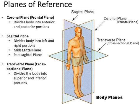
The coronal plane, also known as the frontal plane, is a vertical plane that divides the body into anterior (front) and posterior (back) sections. Imagine a vertical slice through your body from ear to ear; that's the coronal plane. This plane is essential for visualizing structures located on the front and back of the body, such as the relationship between the heart (anterior) and the spinal cord (posterior). Many medical imaging techniques, such as computed tomography (CT) scans and magnetic resonance imaging (MRI) scans, utilize coronal views to assess internal organs and structures.
Clinical Significance of the Coronal Plane: Understanding the coronal plane is paramount in various medical contexts, including:
- Neurological Examinations: Assessing the location and extent of brain injuries or tumors often involves viewing coronal brain slices.
- Orthopedic Assessments: Analyzing fractures, dislocations, or ligament injuries in the limbs frequently requires evaluating coronal views.
- Cardiovascular Imaging: Coronal sections of the heart provide valuable information about the chambers, valves, and surrounding structures.
- Facial Trauma Assessment: Determining the extent of facial bone fractures and soft tissue injuries often utilizes coronal views.
2. Sagittal Plane: Left and Right Division
The sagittal plane is a vertical plane that divides the body into left and right halves. A midsagittal plane, also known as the median plane, divides the body into equal left and right halves. Planes parallel to the midsagittal plane are called parasagittal planes. This plane is vital for examining structures that are paired or symmetrical, such as the lungs, kidneys, or hemispheres of the brain. For example, viewing a sagittal section of the brain reveals the detailed structure of the cerebrum and cerebellum.
3. Transverse Plane (Axial Plane, Horizontal Plane): Superior and Inferior Division
The transverse plane, also called the axial plane or horizontal plane, divides the body into superior (upper) and inferior (lower) sections. Imagine a horizontal slice through your waist; that represents the transverse plane. This plane is crucial for understanding the relationships between different levels of the body, such as the position of abdominal organs relative to the thoracic cavity. Many medical imaging techniques frequently use transverse slices to provide a cross-sectional view of internal structures.
Beyond the Three Primary Planes: Expanding Anatomical Perspective
While the coronal, sagittal, and transverse planes are the primary planes used in anatomical description, understanding additional planes can enhance the precision of anatomical descriptions. These include:
- Oblique Planes: These planes cut the body at any angle that is not parallel to any of the three primary planes. They are often used when a specific structure requires visualization at a unique angle.
- Midclavicular Plane: This vertical plane passes through the midpoints of both clavicles (collarbones). It is frequently used in chest examinations.
- Midaxillary Plane: This vertical plane passes through the midpoints of both axillae (armpits). It's useful for locating structures in the chest and abdomen.
The Importance of Precise Anatomical Terminology
The consistent use of anatomical planes and directional terms is not merely a matter of academic precision; it’s a matter of patient safety and effective communication within the healthcare field. Ambiguity in describing the location of a lesion, injury, or surgical site can have dire consequences. Therefore, a thorough understanding of anatomical planes, combined with accurate directional terminology, is fundamental to medical practice.
Key Directional Terms in Relation to Anatomical Planes:
- Anterior (Ventral): Towards the front of the body.
- Posterior (Dorsal): Towards the back of the body.
- Superior (Cranial): Towards the head.
- Inferior (Caudal): Towards the feet.
- Medial: Towards the midline of the body.
- Lateral: Away from the midline of the body.
- Proximal: Closer to the point of attachment or origin.
- Distal: Further from the point of attachment or origin.
- Superficial: Closer to the surface of the body.
- Deep: Further from the surface of the body.
Applications of Anatomical Planes in Medical Imaging
Medical imaging techniques play a vital role in modern healthcare, providing detailed visualizations of internal body structures. The principles of anatomical planes are fundamental to interpreting these images. Various modalities, such as X-rays, CT scans, MRI scans, and ultrasound, create images that are often viewed in different planes. Understanding the plane of the image is critical for accurately assessing the location and extent of any abnormality.
For instance:
- X-rays: While typically a two-dimensional projection, X-rays can offer valuable information when considering the anatomical plane from which the image is taken.
- CT Scans: CT scans produce cross-sectional images that can be reconstructed in any anatomical plane, providing a comprehensive three-dimensional view. Coronal, sagittal, and axial views are commonly used.
- MRI Scans: Similar to CT scans, MRI scans can generate images in various anatomical planes, offering high-resolution details of soft tissues.
- Ultrasound: Ultrasound images often show structures in a transverse plane, providing a real-time view of organ movement and function.
Clinical Significance: Case Studies and Examples
The practical application of understanding anatomical planes is evident in numerous clinical scenarios. Consider these examples:
- A patient presents with chest pain. A physician might order a chest X-ray or CT scan, examining the coronal plane to assess the position of the heart and lungs and rule out any abnormalities like pneumothorax (collapsed lung) or pericardial effusion (fluid around the heart).
- A patient suffers a head injury. A coronal MRI scan of the brain could reveal the extent of any bleeding or damage to brain tissue.
- A patient experiences knee pain. A coronal MRI scan of the knee joint can help identify ligament tears or meniscus injuries.
- A patient needs surgical intervention for a spinal problem. Preoperative imaging using coronal sections will allow the surgeon to accurately visualize the location and extent of the pathology within the spine.
Conclusion: The Coronal Plane's Indispensable Role in Anatomy and Medicine
The coronal plane, dividing the body into anterior and posterior sections, is a cornerstone of anatomical understanding. Its significance extends far beyond the realm of textbook anatomy; it plays a crucial role in medical diagnosis, treatment planning, and surgical procedures. Understanding this plane, along with other anatomical planes and directional terms, is essential for healthcare professionals and anyone seeking a deeper comprehension of the human body's intricate organization. Mastering this foundational knowledge facilitates clear communication, precise diagnosis, and ultimately, improved patient care. The ability to visualize and interpret images in different anatomical planes is a skill that develops with practice and experience, further highlighting the crucial importance of this concept in medicine and related fields.
Latest Posts
Latest Posts
-
An Acid Is A Proton Donor
Apr 04, 2025
-
Calculate The Heat Capacity Of The Calorimeter
Apr 04, 2025
-
What Is The Correct Order Of Events In Mitosis
Apr 04, 2025
-
Writing A Chemical Equation From A Description Of The Reaction
Apr 04, 2025
-
Highness Or Lowness Of A Sound
Apr 04, 2025
Related Post
Thank you for visiting our website which covers about What Plane Divides The Body Into Anterior And Posterior Parts . We hope the information provided has been useful to you. Feel free to contact us if you have any questions or need further assistance. See you next time and don't miss to bookmark.
