Where Are Chondrocytes And Osteocytes Located
Muz Play
Mar 31, 2025 · 6 min read
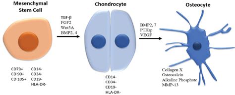
Table of Contents
Where Are Chondrocytes and Osteocytes Located? A Deep Dive into Bone and Cartilage Cells
Understanding the location and function of chondrocytes and osteocytes is crucial to grasping the complexities of the skeletal system. These specialized cells are responsible for the formation, maintenance, and repair of cartilage and bone, respectively. While both contribute to the structural integrity of the body, their locations and roles differ significantly. This article will delve into the precise locations of chondrocytes and osteocytes, exploring their microscopic environments and their broader contribution to skeletal health.
Chondrocytes: The Architects of Cartilage
Chondrocytes are the only cells found in cartilage, a specialized connective tissue that provides flexible support to various parts of the body. Unlike bone, cartilage is avascular, meaning it lacks blood vessels. This unique characteristic impacts nutrient delivery and waste removal, significantly influencing chondrocyte function and location.
Cartilage Types and Chondrocyte Distribution
Cartilage exists in three main types, each with distinct properties and chondrocyte distribution:
1. Hyaline Cartilage: This is the most common type, characterized by its smooth, glassy appearance. It's found in many locations, including:
-
Articular Cartilage: Covering the ends of long bones at joints, enabling smooth, low-friction movement. Chondrocytes here are arranged in isogenous groups, often appearing as clusters within the extracellular matrix. This arrangement reflects their tendency to divide and remain closely associated.
-
Costal Cartilage: Connecting the ribs to the sternum. Chondrocytes in costal cartilage exhibit a more scattered arrangement compared to articular cartilage.
-
Nasal Septum and Tracheal Rings: Providing structural support to these respiratory structures. The chondrocyte distribution in these locations reflects the specific mechanical demands placed on the cartilage.
2. Elastic Cartilage: This type possesses a higher degree of flexibility due to the presence of elastic fibers within the extracellular matrix. Its location includes:
-
Pinna (Auricle) of the Ear: Contributing to the ear's flexible structure. Chondrocytes are dispersed throughout the matrix, reflecting the cartilage's need for flexibility and resilience.
-
Epiglottis: Protecting the airway during swallowing. The chondrocyte arrangement facilitates the epiglottis's ability to bend and adapt to its function.
3. Fibrocartilage: This is the strongest type of cartilage, containing abundant collagen fibers. Its location is primarily in areas subjected to significant compressive forces:
-
Intervertebral Discs: Acting as shock absorbers between vertebrae. Chondrocytes here are arranged in rows parallel to the collagen fibers, aligned with the direction of stress.
-
Menisci of the Knee: Providing stability and shock absorption within the knee joint. The chondrocyte distribution reflects the complex stress patterns experienced by this cartilage.
-
Pubic Symphysis: Connecting the pubic bones of the pelvis. The alignment of chondrocytes reflects the structural demands of this joint.
The Lacunae: Chondrocytes' Microscopic Homes
Chondrocytes reside within small spaces called lacunae (singular: lacuna), which are cavities embedded within the cartilage's extracellular matrix. The matrix itself is a complex network of collagen fibers and ground substance, providing structural support and mediating nutrient exchange for the enclosed chondrocytes. The lacunae provide a protected microenvironment for these cells. The precise shape and arrangement of lacunae and chondrocytes within them vary depending on the cartilage type and location, reflecting the biomechanical forces acting upon the tissue.
Osteocytes: The Guardians of Bone
Osteocytes are the most abundant cells in mature bone tissue, forming a vast interconnected network that plays a critical role in bone remodeling, sensing mechanical stress, and maintaining bone homeostasis. Unlike chondrocytes, osteocytes reside within a highly vascularized and innervated environment.
Bone Tissue Structure and Osteocyte Location
Bone tissue is composed of two main types:
1. Compact Bone: This dense, outer layer of bone provides strength and protection. Osteocytes in compact bone are located within lacunae, similar to chondrocytes but within a much more rigid and mineralized matrix. These lacunae are interconnected by microscopic channels called canaliculi. This intricate network of canaliculi allows for communication and nutrient exchange between osteocytes, ensuring the survival and function of these cells within the dense bone matrix. The canaliculi form a continuous network throughout the bone, facilitating the transport of nutrients and signaling molecules.
2. Spongy (Cancellous) Bone: This less dense, inner layer of bone contains a network of trabeculae (thin, bony plates) and spaces filled with bone marrow. Osteocytes within spongy bone are also located within lacunae, but the arrangement is less organized than in compact bone. The trabeculae provide structural support, and their interconnected nature enhances the overall strength and flexibility of the bone. The proximity of osteocytes to the bone marrow in spongy bone also facilitates communication with other bone cells, such as osteoblasts and osteoclasts.
The Canaliculi: Osteocytes' Communication Network
The canaliculi are essential for osteocyte survival and function. These tiny channels allow for the exchange of nutrients, waste products, and signaling molecules between osteocytes and the blood vessels located in the Haversian canals (central canals) of compact bone. The canalicular system forms a complex network, enabling effective communication and coordination of bone remodeling activities across the entire bone structure. The extensive interconnectedness of the osteocyte network allows for rapid responses to mechanical loading and microdamage, essential for maintaining bone integrity.
Osteocyte Function: Beyond Simple Location
The location of osteocytes within the bone matrix is intimately linked to their function. Their strategic placement allows them to sense mechanical stress and initiate bone remodeling processes to maintain bone strength and adapt to changing demands. They also play a crucial role in regulating mineral homeostasis and coordinating the activities of other bone cells, including osteoblasts (bone-forming cells) and osteoclasts (bone-resorbing cells). The intricate network formed by osteocytes and their canaliculi ensures efficient communication and coordination of these processes, crucial for maintaining the health and integrity of the skeletal system.
Comparing Chondrocytes and Osteocytes: Key Differences
While both chondrocytes and osteocytes are essential for skeletal structure, several key differences highlight their distinct roles:
| Feature | Chondrocytes | Osteocytes |
|---|---|---|
| Location | Cartilage | Bone |
| Vascularity | Avascular (lacks blood vessels) | Vascular (well-supplied with blood vessels) |
| Matrix | Primarily collagen and ground substance | Mineralized matrix (hydroxyapatite crystals) |
| Lacunae | Present, less interconnected | Present, highly interconnected via canaliculi |
| Communication | Primarily via diffusion | Extensive network via canaliculi |
| Major Function | Cartilage formation and maintenance | Bone remodeling, stress sensing, mineral homeostasis |
Conclusion: A Synergistic Relationship
The location of chondrocytes and osteocytes directly reflects their unique functions within the skeletal system. Chondrocytes, nestled within the lacunae of avascular cartilage, contribute to flexible support, while osteocytes, residing within the highly interconnected network of bone tissue, play a vital role in bone remodeling, stress sensing, and maintaining bone health. While their locations and environments differ significantly, both cell types are indispensable for the structural integrity and overall function of the skeleton. The intricate architecture and communication networks within both cartilage and bone, governed by the precise localization of these specialized cells, underline the remarkable complexity and adaptive capacity of the skeletal system.
Latest Posts
Latest Posts
-
Two Different Ionic Compounds Each Contain
Apr 02, 2025
-
Part Ii Equilibria Involving Sparingly Soluble Salts
Apr 02, 2025
-
Adding Strong Acid To A Buffer
Apr 02, 2025
-
The Most Reactive Group In The Periodic Table
Apr 02, 2025
-
How To Write Quadratic Equation From Graph
Apr 02, 2025
Related Post
Thank you for visiting our website which covers about Where Are Chondrocytes And Osteocytes Located . We hope the information provided has been useful to you. Feel free to contact us if you have any questions or need further assistance. See you next time and don't miss to bookmark.
