Are Cilia And Flagella Microtubules Or Microfilaments
Muz Play
Mar 17, 2025 · 6 min read
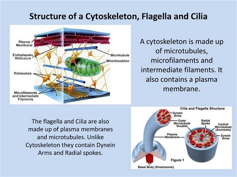
Table of Contents
Are Cilia and Flagella Microtubules or Microfilaments? Understanding the Cellular Machinery of Movement
The mesmerizing, whip-like movements of cilia and flagella are crucial for a vast array of biological processes. From propelling single-celled organisms through liquid environments to facilitating the movement of mucus in our respiratory tracts, these cellular appendages play a vital role in maintaining life as we know it. But what exactly are these structures composed of? Are cilia and flagella microtubules or microfilaments? The answer, as we'll explore in detail, is definitively microtubules. This article delves deep into the structure and function of cilia and flagella, clarifying their composition and highlighting the key differences between microtubules and microfilaments.
The Microtubular Architecture of Cilia and Flagella: A Closer Look
Cilia and flagella, while differing in length and beating pattern, share a strikingly similar internal structure. This conserved architecture is built upon a core scaffolding of microtubules, arranged in a highly organized and characteristic pattern known as the axoneme. The axoneme typically consists of nine outer doublet microtubules surrounding a central pair of singlet microtubules – a 9+2 arrangement.
The 9+2 Arrangement: A Hallmark of Ciliary and Flagellar Structure
This iconic 9+2 arrangement isn't simply an aesthetic feature; it's fundamental to the mechanism of motility. Each doublet microtubule is comprised of a complete A-tubule and a partially complete B-tubule, linked together. These microtubules are not static; they are interconnected by a complex network of proteins, including:
- Nexin: This protein links adjacent doublet microtubules, contributing to the structural integrity of the axoneme.
- Radial spokes: These spokes extend from the A-tubule of each outer doublet to the central pair, playing a critical role in transmitting signals and regulating the dynein arms' activity.
- Dynein arms: These ATPase motor proteins are crucial for generating the movement of cilia and flagella. They are situated on the A-tubule and interact with the B-tubule of the adjacent doublet, causing the microtubules to slide past one another. This sliding action, precisely regulated by the other axoneme components, is the driving force behind ciliary and flagellar beating.
The central pair microtubules are also connected to the outer doublets via a spoke system, further contributing to the coordination of the entire structure. The intricacies of this arrangement ensure the precise, coordinated beating required for effective motility.
Variations in Axoneme Structure: Exceptions to the Rule
While the 9+2 arrangement is the most common, exceptions exist. Some cilia and flagella, particularly in certain species or cell types, exhibit variations in their axoneme structure. For example, some organisms possess cilia or flagella with a 9+0 arrangement, lacking the central pair of microtubules. These variations often reflect functional adaptations to specific environmental or cellular requirements.
Microtubules vs. Microfilaments: Distinguishing Key Features
To fully appreciate why cilia and flagella are composed of microtubules and not microfilaments, it's essential to understand the fundamental differences between these two types of cytoskeletal filaments.
Microtubules: The Pillars of Cellular Organization
Microtubules are long, hollow cylinders formed by the polymerization of α- and β-tubulin dimers. Their rigidity provides structural support to the cell and plays a critical role in various cellular processes, including:
- Intracellular transport: Microtubules serve as tracks for motor proteins like kinesin and dynein, which transport organelles and vesicles throughout the cell.
- Cell division: The mitotic spindle, responsible for segregating chromosomes during cell division, is composed of microtubules.
- Maintaining cell shape: Microtubules contribute to the overall shape and rigidity of the cell.
The dynamic instability of microtubules, their ability to rapidly polymerize and depolymerize, allows for adaptability and responsiveness to cellular signals. This dynamic behavior is particularly important in the regulation of ciliary and flagellar beating.
Microfilaments: The Actin Network for Cell Motility and Structure
Microfilaments, also known as actin filaments, are thinner and more flexible than microtubules. They are composed of polymerized actin monomers and play vital roles in:
- Cell motility: Microfilaments are essential components of the contractile machinery responsible for cell crawling and other forms of movement.
- Cell shape: They contribute to the overall shape and stability of the cell.
- Cytokinesis: They play a critical role in the division of the cytoplasm during cell division.
Unlike the rigid structure of microtubules, microfilaments exhibit a more dynamic and flexible architecture, making them suitable for generating the contractile forces involved in cell motility.
Why Microtubules, Not Microfilaments, in Cilia and Flagella?
The choice of microtubules as the building blocks of cilia and flagella is not arbitrary; it reflects the functional requirements of these appendages. The rigidity and relatively larger diameter of microtubules are crucial for:
- Generating the whip-like motion: The sliding action of microtubules, driven by dynein motor proteins, allows for the large-scale bending movements characteristic of cilia and flagella. The flexibility of microfilaments is insufficient to generate the powerful, coordinated strokes needed for effective propulsion.
- Maintaining structural integrity: The rigid structure of microtubules provides the necessary strength to withstand the forces generated during ciliary and flagellar beating. Microfilaments, being less rigid, would be insufficient to maintain the structural integrity of these long, slender appendages under these stresses.
- Precise coordination of movement: The complex arrangement of microtubules, along with the associated proteins, allows for precise coordination of movement, ensuring efficient propulsion and fluid flow. The simpler organization of microfilaments would be less adept at generating this level of coordinated movement.
The Significance of Cilia and Flagella in Biology
Understanding the structure and function of cilia and flagella extends far beyond basic cell biology. These structures are implicated in a wide range of biological processes and diseases:
Motility in Single-celled Organisms
For many single-celled eukaryotes, cilia and flagella are the primary means of locomotion, enabling them to navigate their environment and find food.
Mucus Clearance in the Respiratory Tract
In humans, the coordinated beating of cilia in the respiratory tract helps to clear mucus and foreign particles from the airways, preventing infection.
Sensory Perception
In some organisms, cilia function as sensory organelles, detecting changes in their environment.
Developmental Processes
Cilia play important roles in embryonic development, guiding cell migration and tissue patterning.
Disease Implications
Disruptions in the structure or function of cilia can lead to various diseases, collectively known as ciliopathies. These conditions can affect a wide range of organ systems, highlighting the crucial role of cilia in human health.
Conclusion: Microtubules as the Foundation of Ciliary and Flagellar Function
In conclusion, cilia and flagella are unequivocally constructed from microtubules, not microfilaments. The unique properties of microtubules – their rigidity, ability to slide past each other, and capacity for complex protein interactions – are essential for generating the coordinated, powerful movements that characterize these cellular appendages. The 9+2 arrangement of microtubules within the axoneme represents a remarkable example of biological engineering, enabling a diverse array of crucial biological functions across a wide spectrum of organisms. Understanding this fundamental aspect of cell biology allows us to appreciate the intricate machinery driving life at its most basic levels and illuminates the mechanisms underlying both health and disease. Further research continues to unravel the complexities of ciliary and flagellar function, leading to advancements in our understanding of cellular processes and disease treatment.
Latest Posts
Latest Posts
-
Is An Atom Smaller Than A Molecule
Mar 17, 2025
-
Why Does Gaining An Electron Give You A Negative Charge
Mar 17, 2025
-
Converting Rectangular Coordinates To Polar Coordinates
Mar 17, 2025
-
How Much Nadh Does Glycolysis Produce
Mar 17, 2025
-
What Is A Principal Agent In Insurance
Mar 17, 2025
Related Post
Thank you for visiting our website which covers about Are Cilia And Flagella Microtubules Or Microfilaments . We hope the information provided has been useful to you. Feel free to contact us if you have any questions or need further assistance. See you next time and don't miss to bookmark.
