Atrioventricular Valves Prevent Backflow Of Blood Into The
Muz Play
Mar 23, 2025 · 6 min read
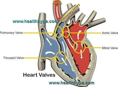
Table of Contents
Atrioventricular Valves: Preventing Backflow and Ensuring Unidirectional Blood Flow
The human heart, a tireless engine of life, relies on a complex system of chambers, vessels, and valves to efficiently pump blood throughout the body. Central to this system are the atrioventricular (AV) valves, crucial structures that prevent the backflow of blood, ensuring unidirectional flow and maintaining the circulatory system's integrity. This article delves into the intricate workings of these valves, exploring their anatomy, physiology, and the critical role they play in maintaining cardiovascular health.
Understanding the Anatomy of Atrioventricular Valves
The heart possesses four chambers: two atria (receiving chambers) and two ventricles (pumping chambers). The atria and ventricles are separated by the AV valves, which act as one-way gates, allowing blood to flow from the atria into the ventricles during diastole (relaxation) but preventing its return during systole (contraction). There are two AV valves:
1. The Tricuspid Valve: Guardian of the Right Atrium-Ventricular Junction
Located between the right atrium and the right ventricle, the tricuspid valve gets its name from its three cusps, or leaflets, which are composed of strong fibrous connective tissue covered by endocardium (the inner lining of the heart). These cusps are connected to papillary muscles via chordae tendineae, strong tendinous cords that prevent the valve leaflets from inverting during ventricular contraction. The tricuspid valve prevents the backflow of deoxygenated blood from the right ventricle into the right atrium.
Key anatomical features of the tricuspid valve:
- Three cusps: Anterior, posterior, and septal.
- Chordae tendineae: Fibrous cords attaching cusps to papillary muscles.
- Papillary muscles: Muscle projections within the right ventricle.
- Annulus fibrosus: A fibrous ring surrounding the valve, providing structural support.
2. The Mitral Valve (Bicuspid Valve): Regulator of Left Atrium-Ventricular Flow
Situated between the left atrium and the left ventricle, the mitral valve, also known as the bicuspid valve due to its two cusps (anterior and posterior), plays a vital role in regulating blood flow from the lungs to the systemic circulation. Similar to the tricuspid valve, the mitral valve’s cusps are connected to papillary muscles via chordae tendineae, preventing valve prolapse (inversion) during ventricular contraction. This prevents the backflow of oxygenated blood from the left ventricle into the left atrium.
Key anatomical features of the mitral valve:
- Two cusps: Anterior and posterior.
- Chordae tendineae: Fibrous cords connecting cusps to papillary muscles.
- Papillary muscles: Muscle projections within the left ventricle.
- Annulus fibrosus: A fibrous ring providing structural support.
The Physiology of Atrioventricular Valves: A Symphony of Opening and Closing
The precise opening and closing of the AV valves are essential for efficient blood flow. This process is entirely passive, driven by pressure gradients across the valves.
Valve Opening: A Matter of Pressure Gradients
During diastole, the atria contract, increasing the atrial pressure. This higher atrial pressure surpasses the ventricular pressure, forcing the AV valves open. Blood then flows freely from the atria into the ventricles.
Valve Closing: Preventing Backflow Through Coordinated Muscle Action
As the ventricles begin to contract during systole, ventricular pressure rapidly increases. This increased ventricular pressure exceeds the atrial pressure, causing the AV valves to close. The chordae tendineae and papillary muscles play a critical role here. As the ventricular pressure rises, the papillary muscles contract, tightening the chordae tendineae and preventing the cusps from inverting or prolapsing into the atria, thereby ensuring a complete closure and preventing backflow.
The Significance of Atrioventricular Valves in Maintaining Cardiovascular Health
The proper functioning of the AV valves is paramount for maintaining cardiovascular health. Any malfunction can lead to serious consequences.
Atrioventricular Valve Diseases: A Spectrum of Cardiovascular Challenges
Several diseases can affect the AV valves, leading to compromised function:
- Mitral Valve Prolapse (MVP): In MVP, one or both mitral valve leaflets bulge back into the left atrium during ventricular contraction. This can cause regurgitation (backflow of blood) and potentially lead to heart failure.
- Mitral Valve Stenosis: Narrowing of the mitral valve opening restricts blood flow from the left atrium to the left ventricle. This reduces cardiac output and can cause shortness of breath and fatigue.
- Tricuspid Regurgitation: The tricuspid valve doesn't close properly, allowing blood to flow back from the right ventricle into the right atrium. This can lead to right-sided heart failure.
- Tricuspid Stenosis: Narrowing of the tricuspid valve opening reduces blood flow from the right atrium to the right ventricle. This can cause symptoms similar to mitral stenosis.
- Atrioventricular Septal Defect (AVSD): This is a congenital heart defect where there's a hole in the wall separating the atria and/or ventricles. This can lead to mixing of oxygenated and deoxygenated blood.
Diagnosing AV Valve Problems: A Multifaceted Approach
Diagnosing AV valve diseases typically involves a combination of methods:
- Physical Examination: Auscultation (listening to the heart sounds with a stethoscope) can reveal murmurs indicative of valve dysfunction.
- Electrocardiogram (ECG): Records the electrical activity of the heart, revealing arrhythmias potentially associated with valve disease.
- Echocardiogram: Utilizes ultrasound to visualize the heart's structure and function, providing detailed images of the valves and assessing their performance.
- Cardiac Catheterization: A more invasive procedure involving inserting a catheter into the heart chambers to measure pressures and assess valve function.
Treatment Strategies for Atrioventricular Valve Disorders
Treatment options vary depending on the severity and type of valve disease:
- Medication: Certain medications can help manage symptoms associated with mild valve dysfunction.
- Valve Repair: Surgical techniques can repair damaged valves, restoring their functionality. This is often preferred over valve replacement when possible, as it preserves the patient's own valve.
- Valve Replacement: In cases of severe valve damage, a damaged valve may need to be replaced with a prosthetic valve, which can be mechanical or biological. Mechanical valves are durable but require lifelong anticoagulation therapy, while biological valves have a limited lifespan but avoid the need for anticoagulants.
Conclusion: The Unsung Heroes of Cardiovascular Health
The atrioventricular valves, often overlooked, are critical components of the circulatory system. Their intricate anatomy and precise physiology ensure unidirectional blood flow, maintaining the heart's efficiency and preventing potentially life-threatening consequences. Understanding their function and the potential for diseases affecting them underscores the importance of regular cardiovascular health checks and prompt medical intervention when necessary. Maintaining a healthy lifestyle, including regular exercise, a balanced diet, and stress management, can contribute to the long-term health of the heart and its vital valves. The AV valves are indeed the unsung heroes of cardiovascular health, silently working to keep our circulatory system functioning optimally. Their continuous and efficient operation is essential for life itself, emphasizing the profound importance of these seemingly simple, yet intricately designed structures. The future of cardiovascular medicine continues to advance, with ongoing research focusing on innovative valve repair and replacement techniques, aiming to improve the lives of individuals affected by AV valve disorders.
Latest Posts
Latest Posts
-
How To Do Inverse Laplace Transforms
Mar 25, 2025
-
Real World Application Of A Linear Equation In 2 Variables
Mar 25, 2025
-
Is Boil A Physical Or Chemical Change
Mar 25, 2025
-
How To Write All Real Numbers In Interval Notation
Mar 25, 2025
-
What Are Rows Called On The Periodic Table
Mar 25, 2025
Related Post
Thank you for visiting our website which covers about Atrioventricular Valves Prevent Backflow Of Blood Into The . We hope the information provided has been useful to you. Feel free to contact us if you have any questions or need further assistance. See you next time and don't miss to bookmark.
