Basic Structure Of The Skin Worksheet Answers
Muz Play
Mar 28, 2025 · 7 min read
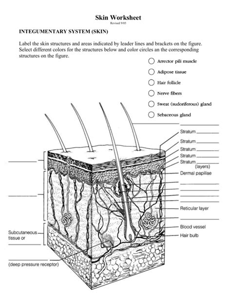
Table of Contents
Basic Structure of the Skin: Worksheet Answers and In-Depth Exploration
Understanding the skin's intricate structure is fundamental to appreciating its vital role in protecting the body. This comprehensive guide provides answers to common worksheet questions on skin structure, delving deeper into each component for a thorough understanding. We'll explore the epidermis, dermis, and hypodermis, examining their individual layers, cell types, and functions. This detailed explanation aims to solidify your knowledge and provide a solid foundation for further study.
The Epidermis: Your Body's Outer Shield
The epidermis, the outermost layer of skin, is a stratified squamous epithelium, meaning it's composed of multiple layers of flattened cells. Its primary function is protection against environmental hazards.
1. Stratum Corneum: The Brick Wall of Dead Cells
This outermost layer is composed of flattened, keratinized dead cells (corneocytes) that are tightly packed together, forming a tough, waterproof barrier. Think of it as a brick wall – strong and resistant to external forces. This layer's primary function is protection against dehydration, abrasion, and pathogen entry. Its thickness varies depending on location; it's thicker on the palms and soles.
2. Stratum Lucidum: A Clear Transition Zone (Only in Thick Skin)
Found only in thick skin (palms, soles), the stratum lucidum is a thin, translucent layer. Its cells are flattened and densely packed with eleidin, a precursor to keratin. It acts as a transitional zone between the stratum granulosum and stratum corneum.
3. Stratum Granulosum: The Granular Layer of Keratin Production
This layer marks the transition from living to dead cells. Cells here contain keratohyalin granules, which contribute to keratin production. Keratinization begins here, hardening the cells and reducing their water content. This process is crucial for the barrier function of the skin.
4. Stratum Spinosum: A Spiky Layer of Interconnected Cells
The stratum spinosum is characterized by its spiny appearance due to the desmosomes, cell junctions that connect the cells. These cells contain numerous melanocytes, which produce melanin, the pigment responsible for skin color and protection against UV radiation. The interconnected nature of this layer provides strength and resilience to the epidermis.
5. Stratum Basale (Germinativum): The Birthplace of New Skin Cells
This deepest layer of the epidermis is where new skin cells are generated through mitosis. It contains keratinocytes, melanocytes, and Merkel cells. Keratinocytes continuously divide and migrate upwards, eventually differentiating into the cells of the more superficial layers. This constant cell renewal ensures the epidermis's ongoing repair and maintenance. Merkel cells are associated with touch sensation, while melanocytes provide vital UV protection.
The Dermis: A Dynamic Layer of Support and Function
The dermis, lying beneath the epidermis, is a much thicker layer of connective tissue. It's responsible for the skin's structural support, elasticity, and numerous functions.
1. Papillary Layer: The Dermal Papillae and Sensory Receptors
This thin, superficial layer of the dermis is characterized by dermal papillae, finger-like projections that interlock with the epidermis. This interdigitation enhances the adhesion between the epidermis and dermis. The papillary layer also contains Meissner's corpuscles, responsible for light touch sensation.
2. Reticular Layer: Strength, Elasticity, and Appendages
The deeper and thicker reticular layer comprises dense irregular connective tissue, providing the skin's strength and elasticity. It contains collagen and elastin fibers, which are crucial for maintaining skin turgor (firmness) and resilience. This layer houses Pacinian corpuscles, which detect deep pressure and vibration. Hair follicles, sebaceous glands (oil glands), and sweat glands (sudoriferous glands) are embedded within the reticular layer.
The Hypodermis: Subcutaneous Fat and Insulation
The hypodermis, or subcutaneous tissue, is the deepest layer of skin. It's primarily composed of adipose tissue (fat cells) and loose connective tissue.
Functions of the Hypodermis:
- Insulation: The fat layer in the hypodermis acts as insulation, protecting the body from temperature fluctuations.
- Energy Storage: Adipose cells store energy in the form of triglycerides.
- Cushioning: The hypodermis cushions and protects underlying organs and structures.
- Attachment: It anchors the skin to underlying muscles and bones.
Skin Appendages: Specialized Structures with Crucial Roles
Several specialized structures are embedded within the dermis and contribute significantly to the skin's overall function.
1. Hair Follicles: Hair Growth and Protection
Hair follicles are tubular invaginations of the epidermis that extend into the dermis. They are responsible for hair growth, offering protection from the sun and physical trauma. The hair follicle cycle involves growth (anagen), regression (catagen), and resting (telogen) phases.
2. Sebaceous Glands: Oil Production and Skin Hydration
Sebaceous glands, associated with hair follicles, produce sebum, an oily substance that lubricates the skin and hair, preventing dryness and cracking. Sebum also has antibacterial properties.
3. Sweat Glands (Sudoriferous Glands): Thermoregulation and Excretion
Sweat glands are responsible for producing sweat, which plays a crucial role in thermoregulation (cooling the body) and excretion of waste products. There are two types of sweat glands: eccrine and apocrine. Eccrine glands are distributed throughout the body and produce a watery sweat, while apocrine glands are located in the axillae and genital areas, producing a thicker, odorous sweat.
Cellular Components: A Closer Look
The skin is composed of various cell types, each contributing to its overall structure and function:
- Keratinocytes: The most abundant cells in the epidermis, responsible for keratin production and forming the protective barrier.
- Melanocytes: Located in the stratum basale, they produce melanin, a pigment that protects against UV radiation. Melanin production varies depending on genetic factors and sun exposure.
- Langerhans Cells: Immune cells that play a role in initiating immune responses against pathogens.
- Merkel Cells: Associated with touch sensation, found in the stratum basale.
- Fibroblasts: Located in the dermis, these cells produce collagen and elastin fibers, contributing to skin strength and elasticity.
Worksheet Answers and Further Elaboration
Let's address some typical worksheet questions concerning the basic structure of the skin:
Q1: Name the three main layers of the skin.
A1: The three main layers of the skin are the epidermis, dermis, and hypodermis (subcutaneous tissue).
Q2: Describe the functions of the epidermis.
A2: The epidermis’s primary function is protection. This includes protection against dehydration, abrasion, UV radiation, and pathogen entry. It also plays a role in maintaining the body's overall homeostasis.
Q3: What is keratinization, and where does it occur?
A3: Keratinization is the process by which keratinocytes produce keratin, a tough, fibrous protein. This process begins in the stratum granulosum and continues as the cells migrate upwards through the epidermis, ultimately forming the protective stratum corneum.
Q4: What are the different layers of the epidermis, and how do they differ?
A4: The epidermis consists of five layers (strata): stratum corneum (dead, keratinized cells), stratum lucidum (only in thick skin, translucent), stratum granulosum (keratinization begins), stratum spinosum (spiny cells, many desmosomes), and stratum basale (actively dividing cells). Each layer has a unique structure and function contributing to the epidermis's overall protective barrier.
Q5: What is the role of the dermis in skin function?
A5: The dermis provides structural support to the skin, maintaining its strength, elasticity, and firmness. It houses blood vessels, nerves, sensory receptors, hair follicles, and glands, contributing to thermoregulation, sensation, and the production of sebum and sweat.
Q6: What are the two main layers of the dermis?
A6: The two main layers of the dermis are the papillary layer (superficial, with dermal papillae and Meissner's corpuscles) and the reticular layer (deeper, with collagen and elastin fibers, Pacinian corpuscles, and appendages).
Q7: What is the hypodermis composed of, and what are its functions?
A7: The hypodermis is primarily composed of adipose tissue (fat cells) and loose connective tissue. Its functions include insulation, energy storage, cushioning, and anchoring the skin to underlying structures.
Q8: Describe the role of melanocytes in protecting the skin.
A8: Melanocytes produce melanin, a pigment that absorbs UV radiation, shielding the underlying cells from its harmful effects. This protection helps prevent sunburns, premature aging, and skin cancer. Melanin production is influenced by genetics and sun exposure.
Q9: What are the functions of sweat glands?
A9: Sweat glands (sudoriferous glands) produce sweat, which contributes to thermoregulation by evaporative cooling. They also play a role in excreting waste products. There are two types: eccrine (watery sweat) and apocrine (thicker, odorous sweat).
Q10: Explain the importance of the skin's barrier function.
A10: The skin's barrier function is critical for protecting the body from external environmental factors such as dehydration, physical trauma, pathogens, and UV radiation. This barrier is primarily maintained by the epidermis's stratified layers and the production of keratin and lipids.
This detailed explanation of the skin's basic structure should provide comprehensive answers to most worksheet questions. Remember to consult additional resources and further research to deepen your understanding. The skin is a complex and fascinating organ with numerous roles in maintaining overall health. Understanding its structure is essential for appreciating its importance and vulnerability.
Latest Posts
Latest Posts
-
Electric Field Of A Charged Disk
Mar 31, 2025
-
5 Blind Man And The Elephant
Mar 31, 2025
-
Which Statement Describes The Citric Acid Cycle
Mar 31, 2025
-
Why Do Plants Love Water In Bio Terms
Mar 31, 2025
-
Identifying The Important Intermolecular Forces In Pure Compounds
Mar 31, 2025
Related Post
Thank you for visiting our website which covers about Basic Structure Of The Skin Worksheet Answers . We hope the information provided has been useful to you. Feel free to contact us if you have any questions or need further assistance. See you next time and don't miss to bookmark.
