Bones Of The Pectoral Girdle And Upper Limb
Muz Play
Mar 24, 2025 · 6 min read
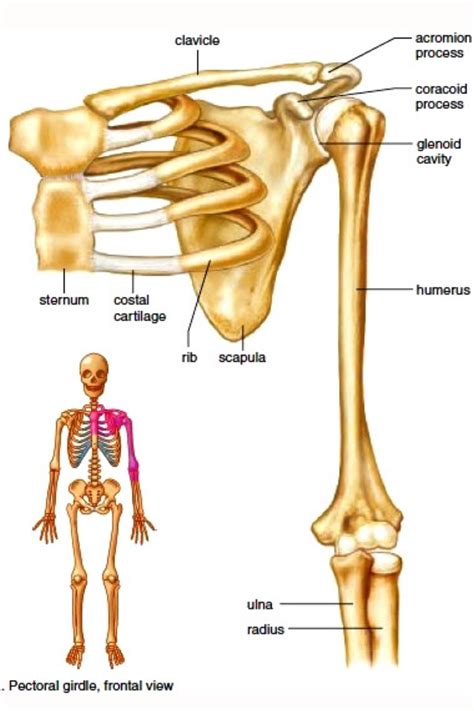
Table of Contents
Bones of the Pectoral Girdle and Upper Limb: A Comprehensive Guide
The human skeletal system is a marvel of engineering, providing structure, support, and protection for our bodies. This article delves deep into the intricate anatomy of the pectoral girdle and upper limb, exploring the individual bones, their articulations, and their crucial roles in movement and function. Understanding this complex system is vital for anyone studying anatomy, physical therapy, orthopedics, or simply anyone interested in the human body's amazing capabilities.
The Pectoral Girdle: The Foundation of Upper Limb Movement
The pectoral girdle, also known as the shoulder girdle, provides the connection between the upper limb and the axial skeleton. Unlike the pelvic girdle, which is firmly attached to the sacrum, the pectoral girdle exhibits greater mobility, contributing to the remarkable range of motion in the upper limb. This mobility, however, comes at the cost of some stability, making the shoulder joint more susceptible to injury.
The pectoral girdle comprises two bones: the clavicle and the scapula.
The Clavicle: The Collarbone's Crucial Role
The clavicle, or collarbone, is a long, S-shaped bone located superficially, easily palpable just above the first rib. Its medial end articulates with the sternum at the sternoclavicular joint, forming the only bony connection between the upper limb and the axial skeleton. The lateral end articulates with the acromion process of the scapula at the acromioclavicular joint.
Key Functions of the Clavicle:
- Transmission of forces: The clavicle acts as a strut, transferring forces from the upper limb to the axial skeleton. This is crucial for activities like pushing, pulling, and weight-bearing.
- Maintaining shoulder stability: It helps to keep the scapula in the correct position, contributing to overall shoulder stability.
- Protection of neurovascular structures: It offers protection to the underlying brachial plexus and subclavian vessels.
The Scapula: The Shoulder Blade's Versatile Design
The scapula, or shoulder blade, is a flat, triangular bone located on the posterior aspect of the thorax. It's remarkably mobile, gliding across the rib cage, allowing for a wide range of arm movements. The scapula possesses several important features:
- Acromion process: The lateral extension of the scapular spine, articulating with the clavicle.
- Coracoid process: A beak-like projection extending anteriorly, serving as an attachment site for several muscles.
- Glenoid cavity: A shallow, pear-shaped fossa that articulates with the head of the humerus, forming the glenohumeral joint (shoulder joint).
- Scapular spine: A prominent ridge running across the posterior surface of the scapula, providing attachment points for muscles.
- Superior, medial, and lateral borders: Define the shape and boundaries of the scapula.
Key Functions of the Scapula:
- Expanding range of motion: Its mobility on the ribcage significantly increases the range of motion of the shoulder joint.
- Muscle attachment: It provides numerous attachment sites for muscles involved in shoulder and arm movements.
- Stabilization of the glenohumeral joint: It plays a significant role in stabilizing the shoulder joint despite its shallow articulation.
The Upper Limb: Bones of the Arm, Forearm, and Hand
The upper limb extends from the shoulder to the fingertips and is remarkably versatile. It's divided into three major segments: the arm, the forearm, and the hand. Each segment contains several bones, contributing to its complexity and functionality.
The Arm: The Humerus - The Long Bone of the Upper Arm
The humerus is the single long bone of the arm, extending from the shoulder to the elbow. Its proximal end articulates with the glenoid cavity of the scapula, forming the glenohumeral joint, and its distal end articulates with the radius and ulna, forming the elbow joint.
Key Features of the Humerus:
- Head: Articulates with the glenoid cavity of the scapula.
- Greater and lesser tubercles: Provide attachment points for rotator cuff muscles.
- Surgical neck: A common site of fracture.
- Deltoid tuberosity: A roughened area providing attachment for the deltoid muscle.
- Capitulum and trochlea: Articulate with the radius and ulna, respectively, at the elbow joint.
- Medial and lateral epicondyles: Provide attachment points for forearm muscles.
The Forearm: Radius and Ulna - Working in Harmony
The forearm comprises two long bones: the radius and the ulna. They articulate with each other at the proximal and distal radioulnar joints, allowing for pronation and supination (rotation of the forearm).
The Radius:
- Head: Articulates with the capitulum of the humerus and the radial notch of the ulna.
- Radial tuberosity: Attachment site for the biceps brachii muscle.
- Styloid process: A pointed projection on the lateral side of the distal end.
The Ulna:
- Olecranon process: Forms the bony prominence of the elbow.
- Trochlear notch: Articulates with the trochlea of the humerus.
- Coronoid process: Participates in the elbow joint articulation.
- Styloid process: A pointed projection on the medial side of the distal end.
Pronation and Supination: The radius and ulna work together to allow the forearm to rotate. Pronation turns the palm downward, while supination turns it upward. This coordinated movement is essential for many daily activities.
The Hand: Carpals, Metacarpals, and Phalanges – A Complex Structure for Dexterity
The hand, the terminal part of the upper limb, is a masterpiece of intricate bone structure, providing dexterity and precision. It's composed of three sets of bones:
-
Carpals: Eight small, carpal bones arranged in two rows. They form the wrist joint and contribute to its complex movements. These bones are often referred to by their location and include the scaphoid, lunate, triquetrum, pisiform, trapezium, trapezoid, capitate, and hamate.
-
Metacarpals: Five long bones forming the palm of the hand. They are numbered I-V, starting from the thumb side. The base of each metacarpal articulates with the carpals, while the head articulates with the phalanges.
-
Phalanges: The bones of the fingers. The thumb (pollex) has two phalanges (proximal and distal), while each of the other fingers (index, middle, ring, and little) has three (proximal, middle, and distal).
Clinical Significance: Fractures and Dislocations
The bones of the pectoral girdle and upper limb are prone to fractures and dislocations due to their involvement in various activities and their relatively exposed positions. Some common injuries include:
- Clavicular fractures: Often occur from direct trauma to the shoulder.
- Scapular fractures: Less common, usually resulting from high-energy trauma.
- Humeral fractures: A common fracture site, especially in the surgical neck and shaft.
- Radial head fractures: Often occur from a fall onto an outstretched hand.
- Colle's fracture: A fracture of the distal radius, commonly seen in falls.
- Scaphoid fractures: A common wrist fracture, often difficult to diagnose.
- Shoulder dislocation: The head of the humerus dislocates from the glenoid cavity, often anteriorly.
- Elbow dislocation: The radius and ulna dislocate from the humerus.
Understanding the anatomy of the pectoral girdle and upper limb is crucial for diagnosing and treating these injuries.
Conclusion: A Complex System, A Remarkable Function
The bones of the pectoral girdle and upper limb represent a sophisticated and integrated system that enables a vast range of movements and functions. From the subtle adjustments of the scapula to the powerful actions of the arm and hand, this system is a testament to the remarkable design of the human body. Through a deeper understanding of the individual bones, their articulations, and their interplay, we gain a profound appreciation for the intricate mechanics that underpin our everyday actions. This knowledge is fundamental for healthcare professionals, athletes, and anyone fascinated by the intricacies of the human form. Further exploration into the muscular attachments and neurological innervation of this region provides an even richer understanding of the complex interplay that defines human movement.
Latest Posts
Latest Posts
-
All Of The Genes Available In A Population
Mar 28, 2025
-
How To Read A Potential Energy Graph
Mar 28, 2025
-
How Many Oxygen Atoms Are In H20
Mar 28, 2025
-
Find Projection Of V Onto U
Mar 28, 2025
-
Moment Of Inertia Of A Stick
Mar 28, 2025
Related Post
Thank you for visiting our website which covers about Bones Of The Pectoral Girdle And Upper Limb . We hope the information provided has been useful to you. Feel free to contact us if you have any questions or need further assistance. See you next time and don't miss to bookmark.
