Bundles Of Axons Within The Central Nervous System Are Called
Muz Play
Mar 20, 2025 · 6 min read
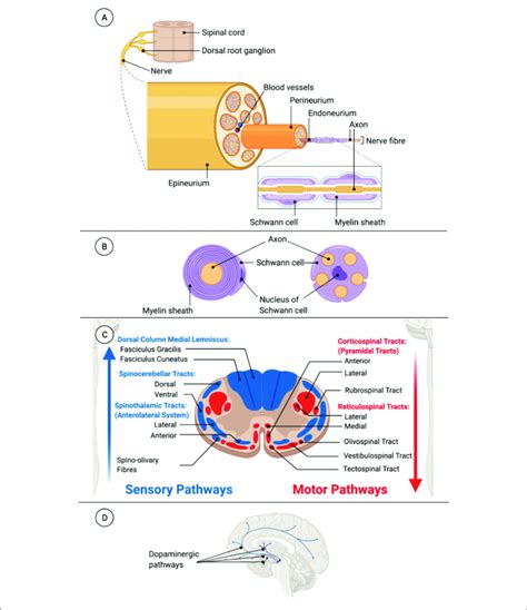
Table of Contents
Bundles of Axons Within the Central Nervous System are Called Tracts: A Deep Dive into Neurological Pathways
The intricate communication network that governs our thoughts, actions, and sensations relies heavily on the efficient transmission of information throughout the central nervous system (CNS). A crucial component of this system is the axon, the long, slender projection of a neuron responsible for transmitting electrical signals. When these axons are bundled together within the CNS, they form structures known as tracts. Understanding the structure, function, and classification of these tracts is fundamental to comprehending the complexity and sophistication of the nervous system.
What are Tracts? A Definition and Clarification
Before delving deeper, let's establish a clear definition. Tracts are bundles of axons located within the central nervous system – specifically, the brain and spinal cord. These bundles are analogous to nerves, which are bundles of axons found in the peripheral nervous system (PNS). However, a key distinction lies in their location and the types of signals they carry. While nerves connect the CNS to peripheral structures like muscles and sensory organs, tracts connect different regions within the CNS itself. This internal connectivity is vital for coordinating various bodily functions and higher-level cognitive processes.
The Anatomy of a Tract: More Than Just Axons
A tract isn't simply a random collection of axons; its structure is highly organized and contributes significantly to its function. While the primary component is, indeed, numerous axons, several other crucial elements contribute to the tract's integrity and efficiency:
1. Myelin Sheath: The Insulation for Speed
Many axons within tracts are covered by a myelin sheath, a fatty insulating layer formed by glial cells. In the CNS, these glial cells are oligodendrocytes. The myelin sheath significantly increases the speed of signal transmission, enabling rapid communication between different brain regions or between the brain and spinal cord. The nodes of Ranvier, gaps in the myelin sheath, further enhance this rapid transmission through saltatory conduction.
2. Neuroglia: Support and Protection
In addition to oligodendrocytes, other glial cells, including astrocytes and microglia, play vital roles within the tract. Astrocytes provide structural support and maintain the extracellular environment, while microglia act as the immune cells of the CNS, protecting against injury and infection. This glial support network is essential for the proper function and survival of the axons within the tract.
3. Connective Tissue: Organization and Structure
While not as prominent as in peripheral nerves, connective tissue elements contribute to the overall organization and structural integrity of tracts. These tissues provide support and help maintain the spatial arrangement of the axons.
Functional Classification of Tracts
Tracts within the CNS can be categorized based on the type of information they transmit. This functional classification is crucial for understanding the specific roles of different tracts in neurological processes.
1. Sensory Tracts (Afferent Tracts): Carrying Sensory Information
These tracts transmit sensory information from the periphery to various areas of the brain. They relay information about touch, pain, temperature, proprioception (sense of body position), and other sensory modalities. Examples include the dorsal column-medial lemniscus pathway, which carries fine touch and proprioception information, and the spinothalamic tract, which transmits pain and temperature signals. The information travels through several relay stations, such as the thalamus, before reaching the final destination in the cerebral cortex for conscious perception.
Key Sensory Tract Examples:
- Spinothalamic Tract: Carries pain, temperature, crude touch, and pressure sensations.
- Dorsal Column-Medial Lemniscus Pathway: Carries fine touch, vibration, and proprioception.
- Spinocerebellar Tracts: Carry proprioceptive information to the cerebellum for coordination and balance.
- Optic Tract: Carries visual information from the retina to the visual cortex.
2. Motor Tracts (Efferent Tracts): Directing Voluntary Movement
Motor tracts convey signals from the brain to muscles, controlling voluntary movements. These tracts originate in the motor cortex and travel down through the brainstem and spinal cord to synapse with motor neurons that directly innervate muscles. The corticospinal tract, also known as the pyramidal tract, is the primary pathway for voluntary movement. Other motor tracts, such as the vestibulospinal tract and reticulospinal tract, play roles in posture, balance, and reflex control.
Key Motor Tract Examples:
- Corticospinal Tract (Pyramidal Tract): Controls voluntary movement of limbs and trunk.
- Rubrospinal Tract: Contributes to motor control, particularly upper limb movement.
- Vestibulospinal Tract: Involved in postural adjustments and balance.
- Reticulospinal Tract: Influences muscle tone and reflexes.
- Tectospinal Tract: Mediates head and eye movements in response to visual stimuli.
3. Association Tracts: Connecting Brain Regions
Association tracts connect different areas within the same hemisphere of the brain. These tracts are crucial for integrating information from various cortical regions, enabling complex cognitive functions like language processing, memory, and problem-solving. They facilitate communication between sensory, motor, and association areas of the cerebral cortex.
Key Association Tract Examples:
- Arcuate Fasciculus: Connects Wernicke's area (language comprehension) and Broca's area (speech production). Damage to this tract can result in conduction aphasia.
- Superior Longitudinal Fasciculus: A major association tract connecting frontal, parietal, temporal, and occipital lobes.
- Uncinate Fasciculus: Connects the frontal and temporal lobes, playing a role in memory and semantic processing.
4. Commissural Tracts: Connecting Hemispheres
Commissural tracts connect corresponding areas in the two hemispheres of the brain, enabling interhemispheric communication. The most prominent commissural tract is the corpus callosum, a large bundle of axons that facilitates the transfer of information between the left and right cerebral hemispheres. This interhemispheric communication is essential for coordinating complex actions and cognitive functions.
Key Commissural Tract Examples:
- Corpus Callosum: The largest commissural tract, connecting the left and right cerebral hemispheres.
- Anterior Commissure: A smaller commissure connecting the temporal lobes and some limbic structures.
- Posterior Commissure: Connects parts of the midbrain and is involved in pupillary light reflexes.
Clinical Significance of Tract Damage
Damage to tracts within the CNS, whether due to injury, disease, or stroke, can have significant consequences, leading to a range of neurological deficits. The specific symptoms depend on the location and extent of the damage.
- Stroke: Disruption of blood supply to a tract can lead to its dysfunction, causing symptoms like paralysis (motor tract damage), sensory loss (sensory tract damage), or cognitive impairment (association tract damage).
- Traumatic Brain Injury (TBI): Physical trauma can cause shearing or tearing of tracts, leading to similar deficits as stroke.
- Multiple Sclerosis (MS): An autoimmune disease targeting myelin, MS can disrupt signal transmission in tracts, causing a wide array of neurological symptoms, including muscle weakness, numbness, vision problems, and cognitive difficulties.
- Tumors: Tumors can compress or invade tracts, leading to focal neurological deficits.
Diagnosing tract damage often involves techniques such as magnetic resonance imaging (MRI) and diffusion tensor imaging (DTI), which allow for visualization of the tracts and identification of lesions or abnormalities.
Conclusion: The Unsung Heroes of Neurological Function
The bundles of axons within the CNS, known as tracts, are far more than just anatomical structures; they are the intricate pathways that underpin our sensory experiences, voluntary movements, and higher-level cognitive abilities. Their highly organized structure, including the myelin sheath and supporting neuroglia, ensures efficient and rapid signal transmission. Understanding the different types of tracts and their functional roles is crucial for comprehending the complexity of the nervous system and the consequences of neurological disorders that affect these vital pathways. Further research into the intricacies of these tracts promises to continue expanding our understanding of the brain and its remarkable capabilities. From the smallest sensory details to the most complex thought processes, the tracts are the silent workhorses that allow the symphony of the nervous system to play out seamlessly.
Latest Posts
Latest Posts
-
Manifest And Latent Functions Of Education
Mar 21, 2025
-
Issuance Of Common Stock Journal Entry
Mar 21, 2025
-
Is Salt Water A Mixture Or Pure Substance
Mar 21, 2025
-
What Is A Change Of State
Mar 21, 2025
-
Integral Of Odd And Even Functions
Mar 21, 2025
Related Post
Thank you for visiting our website which covers about Bundles Of Axons Within The Central Nervous System Are Called . We hope the information provided has been useful to you. Feel free to contact us if you have any questions or need further assistance. See you next time and don't miss to bookmark.
